• 文献检索
  • 文档翻译
  • 深度研究
  • 学术资讯
  • Suppr Zotero 插件Zotero 插件
  • 邀请有礼
  • 套餐&价格
  • 历史记录
应用&插件
Suppr Zotero 插件Zotero 插件浏览器插件Mac 客户端Windows 客户端微信小程序
定价
高级版会员购买积分包购买API积分包
服务
文献检索文档翻译深度研究API 文档MCP 服务
关于我们
关于 Suppr公司介绍联系我们用户协议隐私条款
关注我们

Suppr 超能文献

核心技术专利:CN118964589B侵权必究
粤ICP备2023148730 号-1Suppr @ 2026

文献检索

告别复杂PubMed语法,用中文像聊天一样搜索,搜遍4000万医学文献。AI智能推荐,让科研检索更轻松。

立即免费搜索

文件翻译

保留排版,准确专业,支持PDF/Word/PPT等文件格式,支持 12+语言互译。

免费翻译文档

深度研究

AI帮你快速写综述,25分钟生成高质量综述,智能提取关键信息,辅助科研写作。

立即免费体验

DBA/2J小鼠中进行性神经节细胞丢失和视神经变性是可变的且不对称的。

Progressive ganglion cell loss and optic nerve degeneration in DBA/2J mice is variable and asymmetric.

作者信息

Schlamp Cassandra L, Li Yan, Dietz Joel A, Janssen Katherine T, Nickells Robert W

机构信息

Department of Ophthalmology and Visual Sciences, University of Wisconsin, Madison, WI, USA.

出版信息

BMC Neurosci. 2006 Oct 3;7:66. doi: 10.1186/1471-2202-7-66.

DOI:10.1186/1471-2202-7-66
PMID:17018142
原文链接:https://pmc.ncbi.nlm.nih.gov/articles/PMC1621073/
Abstract

BACKGROUND

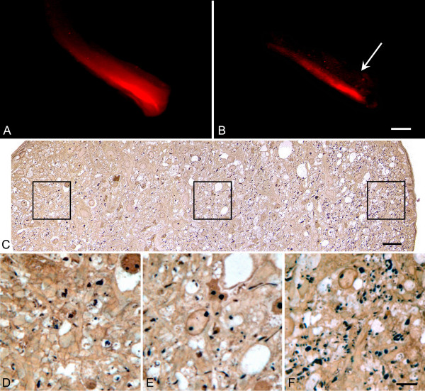
Glaucoma is a chronic neurodegenerative disease of the retina, characterized by the degeneration of axons in the optic nerve and retinal ganglion cell apoptosis. DBA/2J inbred mice develop chronic hereditary glaucoma and are an important model system to study the molecular mechanisms underlying this disease and novel therapeutic interventions designed to attenuate the loss of retinal ganglion cells. Although the genetics of this disease in these mice are well characterized, the etiology of its progression, particularly with respect to retinal degeneration, is not. We have used two separate labeling techniques, post-mortem DiI labeling of axons and ganglion cell-specific expression of the betaGeo reporter gene, to evaluate the time course of optic nerve degeneration and ganglion cell loss, respectively, in aging mice.

RESULTS

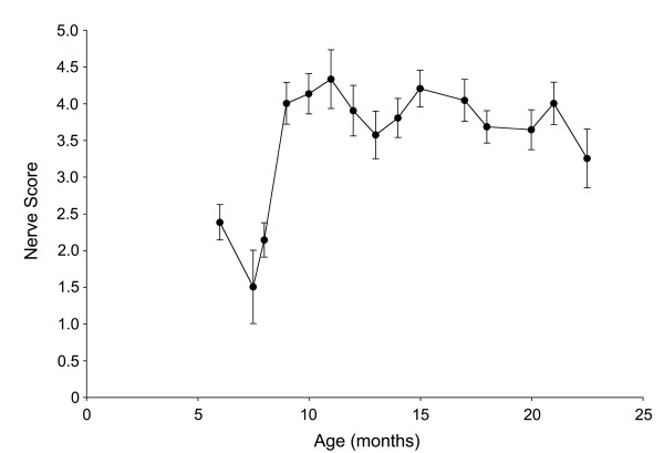
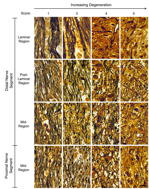
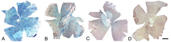
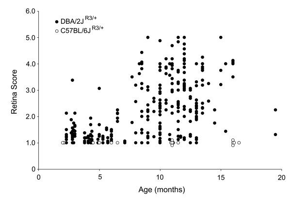
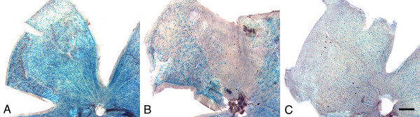
Optic nerve degeneration, characterized by axon loss and gliosis is first apparent in mice between 8 and 9 months of age. Degeneration appears to follow a retrograde course with axons dying from their proximal ends toward the globe. Although nerve damage is typically bilateral, the progression of disease is asymmetric between the eyes of individual mice. Some nerves also exhibit focal preservation of tracts of axons generally in the nasal peripheral region. Ganglion cell loss, as a function of the loss of betaGeo expression, is evident in some mice between 8 and 10 months of age and is prevalent in the majority of mice older than 10.5 months. Most eyes display a uniform loss of ganglion cells throughout the retina, but many younger mice exhibit focal loss of cells in sectors extending from the optic nerve head to the retinal periphery. Similar to what we observe in the optic nerves, ganglion cell loss is often asymmetric between the eyes of the same animal.

CONCLUSION

A comparison of the data collected from the two cohorts of mice used for this study suggests that the initial site of damage in this disease is to the axons in the optic nerve, followed by the subsequent death of the ganglion cell soma.

摘要

背景

青光眼是一种视网膜慢性神经退行性疾病,其特征为视神经轴突退变和视网膜神经节细胞凋亡。DBA/2J近交系小鼠会发生慢性遗传性青光眼,是研究该疾病潜在分子机制以及旨在减轻视网膜神经节细胞损失的新型治疗干预措施的重要模型系统。尽管这些小鼠中该疾病的遗传学特征已得到充分表征,但其进展的病因,尤其是与视网膜退变相关的病因,尚不明确。我们使用了两种不同的标记技术,即轴突的死后DiI标记和βGeo报告基因的神经节细胞特异性表达,分别评估衰老小鼠视神经退变和神经节细胞损失的时间进程。

结果

以轴突损失和胶质增生为特征的视神经退变在8至9月龄小鼠中首次明显出现。退变似乎呈逆行过程,轴突从其近端向眼球方向死亡。尽管神经损伤通常是双侧的,但个体小鼠双眼之间疾病的进展是不对称的。一些神经在鼻周外周区域通常也表现出轴突束的局灶性保留。作为βGeo表达损失的函数,神经节细胞损失在8至10月龄的一些小鼠中明显,在大多数10.5月龄以上的小鼠中普遍存在。大多数眼睛在整个视网膜中显示神经节细胞均匀损失,但许多较年轻的小鼠在从视神经乳头延伸至视网膜周边的扇形区域中表现出细胞的局灶性损失。与我们在视神经中观察到的情况类似,同一动物双眼之间的神经节细胞损失通常也是不对称的。

结论

对本研究中使用的两组小鼠收集的数据进行比较表明,该疾病的初始损伤部位是视神经中的轴突,随后是神经节细胞胞体的死亡。

https://cdn.ncbi.nlm.nih.gov/pmc/blobs/d448/1621073/db5016addccf/1471-2202-7-66-11.jpg
https://cdn.ncbi.nlm.nih.gov/pmc/blobs/d448/1621073/c3fee08d245f/1471-2202-7-66-1.jpg
https://cdn.ncbi.nlm.nih.gov/pmc/blobs/d448/1621073/73d817e8f2c8/1471-2202-7-66-2.jpg
https://cdn.ncbi.nlm.nih.gov/pmc/blobs/d448/1621073/db59ec60f89a/1471-2202-7-66-3.jpg
https://cdn.ncbi.nlm.nih.gov/pmc/blobs/d448/1621073/619f05f1e1d8/1471-2202-7-66-4.jpg
https://cdn.ncbi.nlm.nih.gov/pmc/blobs/d448/1621073/88dd9dcaf269/1471-2202-7-66-5.jpg
https://cdn.ncbi.nlm.nih.gov/pmc/blobs/d448/1621073/8e58d5382db6/1471-2202-7-66-6.jpg
https://cdn.ncbi.nlm.nih.gov/pmc/blobs/d448/1621073/9c251bde6276/1471-2202-7-66-7.jpg
https://cdn.ncbi.nlm.nih.gov/pmc/blobs/d448/1621073/46e062276589/1471-2202-7-66-8.jpg
https://cdn.ncbi.nlm.nih.gov/pmc/blobs/d448/1621073/13c195d0eedd/1471-2202-7-66-9.jpg
https://cdn.ncbi.nlm.nih.gov/pmc/blobs/d448/1621073/a031207bb57e/1471-2202-7-66-10.jpg
https://cdn.ncbi.nlm.nih.gov/pmc/blobs/d448/1621073/db5016addccf/1471-2202-7-66-11.jpg

相似文献

1
Progressive ganglion cell loss and optic nerve degeneration in DBA/2J mice is variable and asymmetric.DBA/2J小鼠中进行性神经节细胞丢失和视神经变性是可变的且不对称的。
BMC Neurosci. 2006 Oct 3;7:66. doi: 10.1186/1471-2202-7-66.
2
Axons of retinal ganglion cells are insulted in the optic nerve early in DBA/2J glaucoma.在DBA/2J青光眼早期,视网膜神经节细胞的轴突在视神经中受到损伤。
J Cell Biol. 2007 Dec 31;179(7):1523-37. doi: 10.1083/jcb.200706181. Epub 2007 Dec 24.
3
Early astrocyte redistribution in the optic nerve precedes axonopathy in the DBA/2J mouse model of glaucoma.在DBA/2J青光眼小鼠模型中,视神经中星形胶质细胞的早期重新分布先于轴突病变。
Exp Eye Res. 2016 Sep;150:22-33. doi: 10.1016/j.exer.2015.11.016. Epub 2015 Dec 2.
4
Glial coverage in the optic nerve expands in proportion to optic axon loss in chronic mouse glaucoma.在慢性小鼠青光眼中,视神经中的神经胶质覆盖范围会随着视神经轴突损失的比例而扩大。
Exp Eye Res. 2016 Sep;150:34-43. doi: 10.1016/j.exer.2016.01.014. Epub 2016 Feb 3.
5
Assessment of inner retina dysfunction and progressive ganglion cell loss in a mouse model of glaucoma.评估青光眼小鼠模型中内视网膜功能障碍和节细胞进行性丧失。
Exp Eye Res. 2014 May;122:40-9. doi: 10.1016/j.exer.2014.02.022. Epub 2014 Mar 12.
6
Retinal ganglion cell degeneration is topological but not cell type specific in DBA/2J mice.在DBA/2J小鼠中,视网膜神经节细胞变性具有拓扑学特征,但并非细胞类型特异性的。
J Cell Biol. 2005 Oct 24;171(2):313-25. doi: 10.1083/jcb.200506099.
7
Reduced retina microglial activation and improved optic nerve integrity with minocycline treatment in the DBA/2J mouse model of glaucoma.在DBA/2J青光眼小鼠模型中,米诺环素治疗可降低视网膜小胶质细胞活化并改善视神经完整性。
Invest Ophthalmol Vis Sci. 2008 Apr;49(4):1437-46. doi: 10.1167/iovs.07-1337.
8
Retinal ganglion cell dendritic atrophy in DBA/2J glaucoma.DBA/2J青光眼患者视网膜神经节细胞树突萎缩
PLoS One. 2013 Aug 19;8(8):e72282. doi: 10.1371/journal.pone.0072282. eCollection 2013.
9
Retinal ganglion cell loss in a rat ocular hypertension model is sectorial and involves early optic nerve axon loss.大鼠眼高压模型中的视网膜神经节细胞丢失呈扇区性,且涉及早期视神经轴突丢失。
Invest Ophthalmol Vis Sci. 2011 Jan 21;52(1):434-41. doi: 10.1167/iovs.10-5856. Print 2011 Jan.
10
Loss of outer retinal neurons and circuitry alterations in the DBA/2J mouse.DBA/2J小鼠中外侧视网膜神经元的丧失及神经回路改变。
Invest Ophthalmol Vis Sci. 2014 Aug 12;55(9):6059-72. doi: 10.1167/iovs.14-14421.

引用本文的文献

1
Morphological Comparison of Astrocytes in the Lamina Cribrosa and Glial Lamina.筛板和神经胶质板中星形胶质细胞的形态学比较。
Invest Ophthalmol Vis Sci. 2025 Mar 3;66(3):1. doi: 10.1167/iovs.66.3.1.
2
Retinal Protection of New Nutraceutical Formulation.新型营养制剂的视网膜保护作用
Pharmaceutics. 2025 Jan 7;17(1):73. doi: 10.3390/pharmaceutics17010073.
3
Preservation of Murine Whole Eyes With Supplemented UW Cold Storage Solution: Anatomical Considerations.补充 UW 冷保存液的小鼠全眼球保存:解剖学考虑。

本文引用的文献

1
Topographic and morphologic analyses of retinal ganglion cell loss in old DBA/2NNia mice.老年DBA/2NNia小鼠视网膜神经节细胞丢失的地形学和形态学分析
Invest Ophthalmol Vis Sci. 2006 May;47(5):1968-74. doi: 10.1167/iovs.05-0955.
2
Inherited glaucoma in DBA/2J mice: pertinent disease features for studying the neurodegeneration.DBA/2J小鼠的遗传性青光眼:用于研究神经退行性变的相关疾病特征。
Vis Neurosci. 2005 Sep-Oct;22(5):637-48. doi: 10.1017/S0952523805225130.
3
Retinal ganglion cell degeneration is topological but not cell type specific in DBA/2J mice.
Transl Vis Sci Technol. 2024 Nov 4;13(11):24. doi: 10.1167/tvst.13.11.24.
4
Proposing a Methodology for Axon-Centric Analysis of IOP-Induced Mechanical Insult.提出一种针对眼压诱导的机械损伤的轴突中心分析方法。
Invest Ophthalmol Vis Sci. 2024 Nov 4;65(13):1. doi: 10.1167/iovs.65.13.1.
5
Morphological comparison of astrocytes in the lamina cribrosa and glial lamina.筛板和神经胶质板中星形胶质细胞的形态学比较。
bioRxiv. 2024 Sep 10:2024.09.07.610493. doi: 10.1101/2024.09.07.610493.
6
Differential protection by nicotinamide in a mouse model of glaucoma DBA/2J revealed by second-harmonic generation microscopy.二倍频产生显微镜观察到烟酰胺对 DBA/2J 型青光眼小鼠的差异保护作用。
PLoS One. 2024 Sep 10;19(9):e0309400. doi: 10.1371/journal.pone.0309400. eCollection 2024.
7
Improved MRI methods to quantify retinal and choroidal blood flow applied to a model of glaucoma.应用于青光眼模型的用于量化视网膜和脉络膜血流的改进磁共振成像方法。
Front Ophthalmol (Lausanne). 2024 May 13;4:1385495. doi: 10.3389/fopht.2024.1385495. eCollection 2024.
8
Differential Retinal Ganglion Cell Vulnerability, A Critical Clue for the Identification of Neuroprotective Genes in Glaucoma.视网膜神经节细胞的差异性易损性:青光眼神经保护基因鉴定的关键线索
Front Ophthalmol (Lausanne). 2022 May 31;2:905352. doi: 10.3389/fopht.2022.905352. eCollection 2022.
9
Sustained Retinal Defocus Increases the Effect of Induced Myopia on the Retinal Astrocyte Template.持续视网膜离焦增加诱导性近视对视网膜星形胶质细胞模板的影响。
Cells. 2024 Mar 29;13(7):595. doi: 10.3390/cells13070595.
10
Protective effects of nicotinamide in a mouse model of glaucoma DBA/2 studied by second-harmonic generation microscopy.通过二次谐波产生显微镜研究烟酰胺在青光眼DBA/2小鼠模型中的保护作用。
bioRxiv. 2024 Apr 18:2024.03.07.583928. doi: 10.1101/2024.03.07.583928.
在DBA/2J小鼠中,视网膜神经节细胞变性具有拓扑学特征,但并非细胞类型特异性的。
J Cell Biol. 2005 Oct 24;171(2):313-25. doi: 10.1083/jcb.200506099.
4
Susceptibility to neurodegeneration in a glaucoma is modified by Bax gene dosage.青光眼患者对神经退行性变的易感性受Bax基因剂量的影响。
PLoS Genet. 2005 Jul;1(1):17-26. doi: 10.1371/journal.pgen.0010004. Epub 2005 Jul 25.
5
Changes in retinal neuronal populations in the DBA/2J mouse.DBA/2J小鼠视网膜神经元群体的变化。
Cell Tissue Res. 2005 Apr;320(1):51-9. doi: 10.1007/s00441-004-1062-8. Epub 2005 Feb 16.
6
The progressive nature of Wallerian degeneration in wild-type and slow Wallerian degeneration (WldS) nerves.野生型和慢沃勒变性(WldS)神经中沃勒变性的进行性特征。
BMC Neurosci. 2005 Feb 1;6:6. doi: 10.1186/1471-2202-6-6.
7
Regional optic nerve damage in experimental mouse glaucoma.实验性小鼠青光眼的局部视神经损伤
Invest Ophthalmol Vis Sci. 2004 Dec;45(12):4352-8. doi: 10.1167/iovs.04-0355.
8
Insertion of the beta Geo promoter trap into the Fem1c gene of ROSA3 mice.将βGeo启动子陷阱插入ROSA3小鼠的Fem1c基因中。
Mol Cell Biol. 2004 May;24(9):3794-803. doi: 10.1128/MCB.24.9.3794-3803.2004.
9
Interocular asymmetry of visual field defects in primary open angle glaucoma and primary angle-closure glaucoma.原发性开角型青光眼和原发性闭角型青光眼视野缺损的双眼不对称性
Eye (Lond). 2004 Apr;18(4):365-8. doi: 10.1038/sj.eye.6700664.
10
Retinal neurodegeneration in the DBA/2J mouse-a model for ocular hypertension.DBA/2J小鼠的视网膜神经变性——一种高眼压模型
Acta Neuropathol. 2004 Apr;107(4):352-8. doi: 10.1007/s00401-003-0816-9. Epub 2004 Jan 24.