• 文献检索
  • 文档翻译
  • 深度研究
  • 学术资讯
  • Suppr Zotero 插件Zotero 插件
  • 邀请有礼
  • 套餐&价格
  • 历史记录
应用&插件
Suppr Zotero 插件Zotero 插件浏览器插件Mac 客户端Windows 客户端微信小程序
定价
高级版会员购买积分包购买API积分包
服务
文献检索文档翻译深度研究API 文档MCP 服务
关于我们
关于 Suppr公司介绍联系我们用户协议隐私条款
关注我们

Suppr 超能文献

核心技术专利:CN118964589B侵权必究
粤ICP备2023148730 号-1Suppr @ 2026

文献检索

告别复杂PubMed语法,用中文像聊天一样搜索,搜遍4000万医学文献。AI智能推荐,让科研检索更轻松。

立即免费搜索

文件翻译

保留排版,准确专业,支持PDF/Word/PPT等文件格式,支持 12+语言互译。

免费翻译文档

深度研究

AI帮你快速写综述,25分钟生成高质量综述,智能提取关键信息,辅助科研写作。

立即免费体验

治疗引起的骨髓变化的磁共振成像

MR imaging of therapy-induced changes of bone marrow.

作者信息

Daldrup-Link Heike E, Henning Tobias, Link Thomas M

机构信息

Department of Radiology, University of California San Francisco, 505 Parnassus Ave., San Francisco, CA, 94143-0628, USA.

出版信息

Eur Radiol. 2007 Mar;17(3):743-61. doi: 10.1007/s00330-006-0404-1. Epub 2006 Sep 21.

DOI:10.1007/s00330-006-0404-1
PMID:17021706
原文链接:https://pmc.ncbi.nlm.nih.gov/articles/PMC1797072/
Abstract

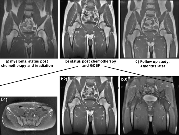
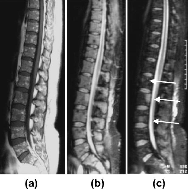
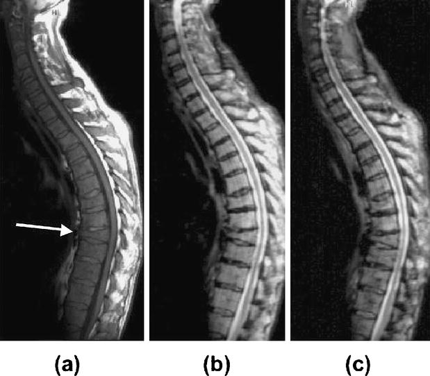
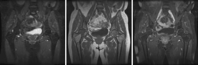
MR imaging of bone marrow infiltration by hematologic malignancies provides non-invasive assays of bone marrow cellularity and vascularity to supplement the information provided by bone marrow biopsies. This article will review the MR imaging findings of bone marrow infiltration by hematologic malignancies with special focus on treatment effects. MR imaging findings of the bone marrow after radiation therapy and chemotherapy will be described. In addition, changes in bone marrow microcirculation and metabolism after anti-angiogenesis treatment will be reviewed. Finally, new specific imaging techniques for the depiction of regulatory events that control blood vessel growth and cell proliferation will be discussed. Future developments are directed to yield comprehensive information about bone marrow structure, function and microenvironment.

摘要

血液系统恶性肿瘤骨髓浸润的磁共振成像(MR)可为骨髓细胞密度和血管分布提供非侵入性检测,以补充骨髓活检所提供的信息。本文将回顾血液系统恶性肿瘤骨髓浸润的MR成像表现,特别关注治疗效果。将描述放疗和化疗后骨髓的MR成像表现。此外,还将回顾抗血管生成治疗后骨髓微循环和代谢的变化。最后,将讨论用于描绘控制血管生长和细胞增殖的调节事件的新的特异性成像技术。未来的发展方向是获取有关骨髓结构、功能和微环境的全面信息。

https://cdn.ncbi.nlm.nih.gov/pmc/blobs/9e14/1797072/c11275c4f77b/330_2006_404_Fig12_HTML.jpg
https://cdn.ncbi.nlm.nih.gov/pmc/blobs/9e14/1797072/5465351e5190/330_2006_404_Fig1_HTML.jpg
https://cdn.ncbi.nlm.nih.gov/pmc/blobs/9e14/1797072/05a6741daf62/330_2006_404_Fig2_HTML.jpg
https://cdn.ncbi.nlm.nih.gov/pmc/blobs/9e14/1797072/1c0dd2a124b6/330_2006_404_Fig3_HTML.jpg
https://cdn.ncbi.nlm.nih.gov/pmc/blobs/9e14/1797072/d7275151e84a/330_2006_404_Fig4_HTML.jpg
https://cdn.ncbi.nlm.nih.gov/pmc/blobs/9e14/1797072/e0b47e123405/330_2006_404_Fig5_HTML.jpg
https://cdn.ncbi.nlm.nih.gov/pmc/blobs/9e14/1797072/ad573cdbefcc/330_2006_404_Fig6_HTML.jpg
https://cdn.ncbi.nlm.nih.gov/pmc/blobs/9e14/1797072/46c4df6254d8/330_2006_404_Fig7_HTML.jpg
https://cdn.ncbi.nlm.nih.gov/pmc/blobs/9e14/1797072/fe6d552c1ce7/330_2006_404_Fig8_HTML.jpg
https://cdn.ncbi.nlm.nih.gov/pmc/blobs/9e14/1797072/40fb65e01ed1/330_2006_404_Fig9_HTML.jpg
https://cdn.ncbi.nlm.nih.gov/pmc/blobs/9e14/1797072/ccd0958cfbe1/330_2006_404_Fig10_HTML.jpg
https://cdn.ncbi.nlm.nih.gov/pmc/blobs/9e14/1797072/d68472878ee6/330_2006_404_Fig11_HTML.jpg
https://cdn.ncbi.nlm.nih.gov/pmc/blobs/9e14/1797072/c11275c4f77b/330_2006_404_Fig12_HTML.jpg

相似文献

1
MR imaging of therapy-induced changes of bone marrow.治疗引起的骨髓变化的磁共振成像
Eur Radiol. 2007 Mar;17(3):743-61. doi: 10.1007/s00330-006-0404-1. Epub 2006 Sep 21.
2
[Diagnosis of bone marrow disorders: is MRT sufficient?].[骨髓疾病的诊断:磁共振成像是否足够?]
Radiologe. 2007 Aug;47(8):660-2. doi: 10.1007/s00117-007-1541-5.
3
New perspectives on bone marrow contrast agents and molecular imaging.骨髓造影剂与分子成像的新视角
Semin Musculoskelet Radiol. 2009 Jun;13(2):145-56. doi: 10.1055/s-0029-1220885. Epub 2009 May 19.
4
MRI of bone marrow abnormalities in hematological malignancies.血液恶性肿瘤骨髓异常的 MRI 表现。
Diagn Interv Radiol. 2013 Sep-Oct;19(5):393-9. doi: 10.5152/dir.2013.067.
5
Therapy Effect: Impact on Bone Marrow Morphology.治疗效果:对骨髓形态的影响。
Surg Pathol Clin. 2016 Mar;9(1):177-87. doi: 10.1016/j.path.2015.09.006.
6
Magnetic resonance imaging of the bone marrow in hematologic malignancies.血液系统恶性肿瘤的骨髓磁共振成像
Blood. 1997 Sep 15;90(6):2127-47.
7
Tumor infiltration of bone marrow in patients with hemato-logical malignancies: dynamic contrast-enhanced magnetic resonance imaging.血液系统恶性肿瘤患者骨髓的肿瘤浸润:动态对比增强磁共振成像
Chin Med J (Engl). 2006 Aug 5;119(15):1256-62.
8
Magnetic resonance imaging of bone marrow in oncology, Part 2.肿瘤学中骨髓的磁共振成像,第2部分。
Skeletal Radiol. 2007 Nov;36(11):1017-27. doi: 10.1007/s00256-007-0308-4. Epub 2007 May 11.
9
[Angiogenesis in patients with hematologic malignancies].[血液系统恶性肿瘤患者的血管生成]
Onkologie. 2001 Sep;24 Suppl 5:75-80. doi: 10.1159/000055192.
10
Magnetic resonance imaging of bone marrow in hematologic malignancies.血液系统恶性肿瘤的骨髓磁共振成像
Int J Hematol. 1997 Dec;66(4):413-22. doi: 10.1016/s0925-5710(97)00085-6.

引用本文的文献

1
Imaging paediatric bone marrow in immunocompromised patients.免疫功能低下患者的儿科骨髓成像
Pediatr Radiol. 2025 Apr;55(4):792-805. doi: 10.1007/s00247-024-06153-7. Epub 2025 Jan 23.
2
Visual assessment and quantitative analysis of dual-energy CT virtual non-calcium in imaging diagnosis of multiple myeloma.双能CT虚拟去钙成像在多发性骨髓瘤诊断中的视觉评估与定量分析
Skeletal Radiol. 2025 May;54(5):1059-1070. doi: 10.1007/s00256-024-04805-1. Epub 2024 Oct 8.
3
Bone marrow edema in children: chronic nonbacterial osteomyelitis and its mimickers.

本文引用的文献

1
Performance test of an LSO-APD detector in a 7-T MRI scanner for simultaneous PET/MRI.用于同时进行PET/MRI的LSO-APD探测器在7-T MRI扫描仪中的性能测试。
J Nucl Med. 2006 Apr;47(4):639-47.
2
Whole-body MRI in the detection of bone marrow infiltration in patients with plasma cell neoplasms in comparison to the radiological skeletal survey.与放射学骨骼检查相比,全身MRI在浆细胞肿瘤患者骨髓浸润检测中的应用
Eur Radiol. 2006 May;16(5):1005-14. doi: 10.1007/s00330-005-0055-7. Epub 2006 Feb 4.
3
Ferumoxtran-10-enhanced MR imaging of the bone marrow before and after conditioning therapy in patients with non-Hodgkin lymphomas.
儿童骨髓水肿:慢性非细菌性骨髓炎及其模仿者
Ther Adv Musculoskelet Dis. 2024 Sep 18;16:1759720X241278438. doi: 10.1177/1759720X241278438. eCollection 2024.
4
From guidelines to radiology practice: navigating the 2023 ASCO guidelines for advanced gastroesophageal cancer and beyond.从指南到放射学实践:解读2023年美国临床肿瘤学会晚期胃食管癌及其他相关疾病指南
Abdom Radiol (NY). 2025 Jan;50(1):78-93. doi: 10.1007/s00261-024-04499-y. Epub 2024 Aug 9.
5
Does the presence of macroscopic intralesional fat exclude malignancy? An analysis of 613 histologically proven malignant bone lesions.宏观瘤内脂肪的存在是否排除恶性肿瘤?613 例组织学证实的恶性骨病变的分析。
Eur Radiol. 2024 Oct;34(10):6581-6589. doi: 10.1007/s00330-024-10687-7. Epub 2024 Mar 15.
6
False Liver Metastasis by Positron Emission Tomography/Computed Tomography Scan after Chemoradiotherapy for Esophageal Cancer-Potential Overstaged Pitfalls of Treatment.食管癌放化疗后正电子发射断层扫描/计算机断层扫描显示的假肝转移——治疗中潜在的分期过度陷阱
Cancers (Basel). 2024 Feb 26;16(5):948. doi: 10.3390/cancers16050948.
7
MRI Methods to Visualize and Quantify Adipose Tissue in Health and Disease.用于可视化和量化健康与疾病状态下脂肪组织的MRI方法
Biomedicines. 2023 Nov 29;11(12):3179. doi: 10.3390/biomedicines11123179.
8
Detection of bone marrow metastases in children and young adults with solid cancers with diffusion-weighted MRI.应用弥散加权 MRI 检测儿童和青年实体瘤患者骨髓转移。
Skeletal Radiol. 2023 Jun;52(6):1179-1192. doi: 10.1007/s00256-022-04240-0. Epub 2022 Nov 28.
9
Study of bone marrow microstructure in healthy young adults using intravoxel incoherent motion diffusion-weighted MRI.使用体素内不相干运动扩散加权 MRI 研究健康年轻成年人的骨髓微观结构。
Front Endocrinol (Lausanne). 2022 Nov 11;13:958151. doi: 10.3389/fendo.2022.958151. eCollection 2022.
10
Dual-energy CT based monitoring of treatment-induced bone marrow changes in lung cancer patients: preliminary results.基于双能CT监测肺癌患者治疗引起的骨髓变化:初步结果
Quant Imaging Med Surg. 2022 Mar;12(3):1871-1881. doi: 10.21037/qims-21-545.
非霍奇金淋巴瘤患者预处理治疗前后的骨髓钆布醇-10增强磁共振成像
Eur Radiol. 2006 Mar;16(3):598-607. doi: 10.1007/s00330-005-0045-9. Epub 2005 Nov 12.
4
Ultrasmall superparamagnetic iron-oxide-enhanced MR imaging of normal bone marrow in rodents: original research original research.超小型超顺磁性氧化铁增强的啮齿动物正常骨髓磁共振成像:原始研究 原始研究
Acad Radiol. 2005 Sep;12(9):1190-7. doi: 10.1016/j.acra.2005.05.014.
5
[Correlation of MRI and histopathology of bone marrow in patients with multiple myeloma].[多发性骨髓瘤患者骨髓的MRI与组织病理学相关性]
Rofo. 2005 Aug;177(8):1116-22. doi: 10.1055/s-2005-858362.
6
Dynamic contrast-enhanced MRI for assessing the disease activity of multiple myeloma: a comparative study with histology and clinical markers.动态对比增强磁共振成像评估多发性骨髓瘤疾病活动度:与组织学及临床标志物的对比研究
J Magn Reson Imaging. 2005 Jul;22(1):154-62. doi: 10.1002/jmri.20349.
7
Normal variants and frequent marrow alterations that simulate bone marrow lesions at MR imaging.正常变异及在磁共振成像中模拟骨髓病变的常见骨髓改变。
Radiol Clin North Am. 2005 Jul;43(4):761-70, ix. doi: 10.1016/j.rcl.2005.01.007.
8
Lymphoma: diagnosis, staging, natural history, and treatment strategies.淋巴瘤:诊断、分期、自然史及治疗策略。
Semin Oncol. 2005 Feb;32(1 Suppl 1):S4-10. doi: 10.1053/j.seminoncol.2005.01.008.
9
Postirradiation sarcoma: a case report and current review.放疗后肉瘤:一例病例报告及当前综述
Am J Otolaryngol. 2005 Jan-Feb;26(1):71-4. doi: 10.1016/j.amjoto.2004.08.005.
10
Imaging therapeutic response in human bone marrow using rapid whole-body MRI.利用快速全身磁共振成像对人类骨髓中的治疗反应进行成像。
Magn Reson Med. 2004 Dec;52(6):1234-8. doi: 10.1002/mrm.20291.