Letessier Anne, Sircoulomb Fabrice, Ginestier Christophe, Cervera Nathalie, Monville Florence, Gelsi-Boyer Véronique, Esterni Benjamin, Geneix Jeannine, Finetti Pascal, Zemmour Christophe, Viens Patrice, Charafe-Jauffret Emmanuelle, Jacquemier Jocelyne, Birnbaum Daniel, Chaffanet Max
Centre de Recherche en Cancérologie de Marseille, Département d'Oncologie Moléculaire, UMR599 Inserm/Institut Paoli-Calmettes, Marseille, France.
BMC Cancer. 2006 Oct 13;6:245. doi: 10.1186/1471-2407-6-245.
Oncogene amplification and overexpression occur in tumor cells. Amplification status may provide diagnostic and prognostic information and may lead to new treatment strategies. Chromosomal regions 8p12, 8q24, 11q13, 17q12 and 20q13 are recurrently amplified in breast cancers.
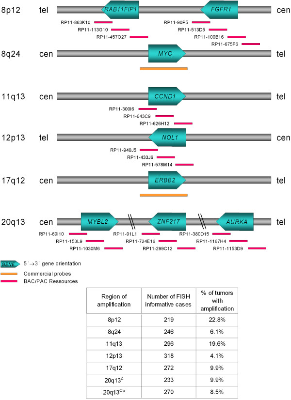
To assess the frequencies and clinical impact of amplifications, we analyzed 547 invasive breast tumors organized in a tissue microarray (TMA) by fluorescence in situ hybridization (FISH) and calculated correlations with histoclinical features and prognosis. BAC probes were designed for: (i) two 8p12 subregions centered on RAB11FIP1 and FGFR1 loci, respectively; (ii) 11q13 region centered on CCND1; (iii) 12p13 region spanning NOL1; and (iv) three 20q13 subregions centered on MYBL2, ZNF217 and AURKA, respectively. Regions 8q24 and 17q12 were analyzed with MYC and ERBB2 commercial probes, respectively.
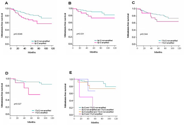
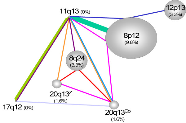
We observed amplification of 8p12 (amplified at RAB11FIP1 and/or FGFR1) in 22.8%, 8q24 in 6.1%, 11q13 in 19.6%, 12p13 in 4.1%, 17q12 in 9.9%, 20q13Z (amplified at ZNF217 only) in 9.9%, and 20q13Co (co-amplification of two or three 20q13 loci) in 8.5% of cases. The 8q24, 12p13, and 17q12 amplifications were correlated with high grade. The most frequent single amplifications were 8p12 (9.8%), 8q24 (3.3%) and 12p13 (3.3%), 20q13Z and 20q13Co (1.6%) regions. The 17q12 and 11q13 regions were never found amplified alone. The most frequent co-amplification was 8p12/11q13. Amplifications of 8p12 and 17q12 were associated with poor outcome. Amplification of 12p13 was associated with basal molecular subtype.
Our results establish the frequencies, prognostic impacts and subtype associations of various amplifications and co-amplifications in breast cancers.
癌基因扩增和过表达发生于肿瘤细胞中。扩增状态可提供诊断和预后信息,并可能带来新的治疗策略。染色体区域8p12、8q24、11q13、17q12和20q13在乳腺癌中经常发生扩增。
为评估扩增的频率及其临床影响,我们通过荧光原位杂交(FISH)分析了组织微阵列(TMA)中547例浸润性乳腺肿瘤,并计算其与组织临床特征及预后的相关性。设计了BAC探针用于:(i)分别以RAB11FIP1和FGFR1基因为中心的两个8p12亚区域;(ii)以CCND1为中心的11q13区域;(iii)跨越NOL1的12p13区域;(iv)分别以MYBL2、ZNF217和AURKA为中心的三个20q13亚区域。8q24和17q12区域分别使用MYC和ERBB2商业探针进行分析。
我们观察到,22.8%的病例中存在8p12(在RAB11FIP1和/或FGFR1处扩增),6.1%的病例中存在8q24,19.6%的病例中存在11q13,4.1%的病例中存在12p13,9.9%的病例中存在17q12,9.9%的病例中存在20q13Z(仅在ZNF217处扩增),8.5%的病例中存在20q13Co(两个或三个20q13位点共扩增)。8q24、12p13和17q12扩增与高级别相关联。最常见的单个扩增区域为8p12(9.8%)、8q24(3.3%)和12p13(3.3%)、20q13Z和20q13Co(1.6%)区域。从未发现17q12和11q13区域单独扩增。最常见的共扩增为8p12/11q13。8p12和17q12扩增与不良预后相关。12p13扩增与基底分子亚型相关。
我们的结果确定了乳腺癌中各种扩增和共扩增的频率、预后影响及亚型关联。