• 文献检索
  • 文档翻译
  • 深度研究
  • 学术资讯
  • Suppr Zotero 插件Zotero 插件
  • 邀请有礼
  • 套餐&价格
  • 历史记录
应用&插件
Suppr Zotero 插件Zotero 插件浏览器插件Mac 客户端Windows 客户端微信小程序
定价
高级版会员购买积分包购买API积分包
服务
文献检索文档翻译深度研究API 文档MCP 服务
关于我们
关于 Suppr公司介绍联系我们用户协议隐私条款
关注我们

Suppr 超能文献

核心技术专利:CN118964589B侵权必究
粤ICP备2023148730 号-1Suppr @ 2026

文献检索

告别复杂PubMed语法,用中文像聊天一样搜索,搜遍4000万医学文献。AI智能推荐,让科研检索更轻松。

立即免费搜索

文件翻译

保留排版,准确专业,支持PDF/Word/PPT等文件格式,支持 12+语言互译。

免费翻译文档

深度研究

AI帮你快速写综述,25分钟生成高质量综述,智能提取关键信息,辅助科研写作。

立即免费体验

使用二维凝胶电泳对预定义蛋白质组进行标准化及表达变化分析:一项利用差异凝胶电泳(DIGE)技术对帕金森病动物模型中左旋多巴诱导的异动症进行蛋白质组学研究。

Normalization and expression changes in predefined sets of proteins using 2D gel electrophoresis: a proteomic study of L-DOPA induced dyskinesia in an animal model of Parkinson's disease using DIGE.

作者信息

Kultima Kim, Scholz Birger, Alm Henrik, Sköld Karl, Svensson Marcus, Crossman Alan R, Bezard Erwan, Andrén Per E, Lönnstedt Ingrid

机构信息

Department of Pharmaceutical Biosciences, Division of Toxicology, Uppsala University, BMC, Box 594, SE-75124 Uppsala, Sweden.

出版信息

BMC Bioinformatics. 2006 Oct 26;7:475. doi: 10.1186/1471-2105-7-475.

DOI:10.1186/1471-2105-7-475
PMID:17067368
原文链接:https://pmc.ncbi.nlm.nih.gov/articles/PMC1635739/
Abstract

BACKGROUND

Two-Dimensional Difference In Gel Electrophoresis (2D-DIGE) is a powerful tool for measuring differences in protein expression between samples or conditions. However, to remove systematic variability within and between gels the data has to be normalized. In this study we examined the ability of four existing and four novel normalization methods to remove systematic bias in data produced with 2D-DIGE. We also propose a modification of an existing method where the statistical framework determines whether a set of proteins shows an association with the predefined phenotypes of interest. This method was applied to our data generated from a monkey model (Macaca fascicularis) of Parkinson's disease.

RESULTS

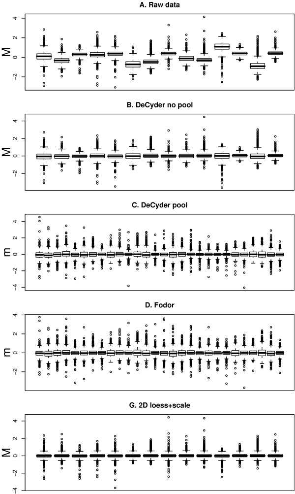
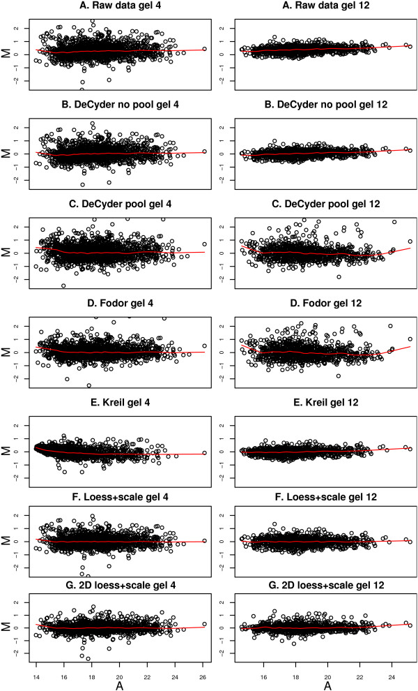
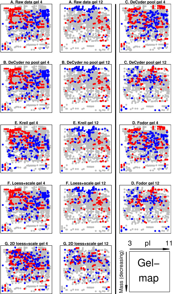
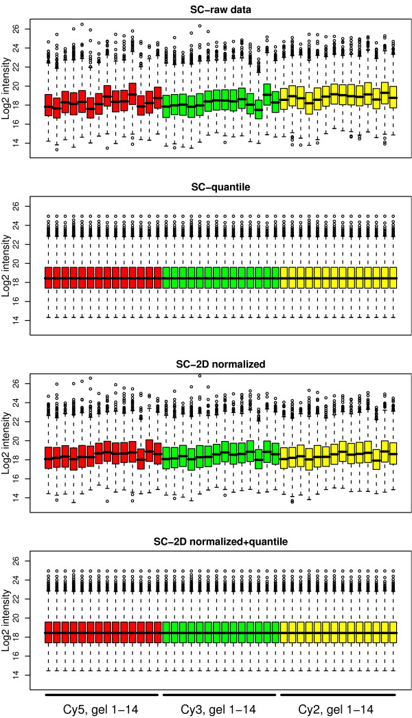
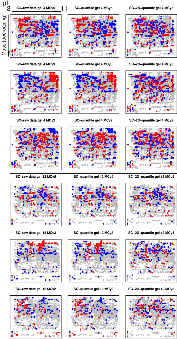
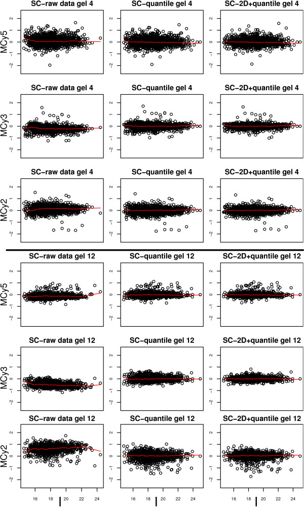
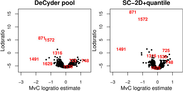
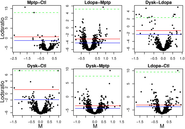
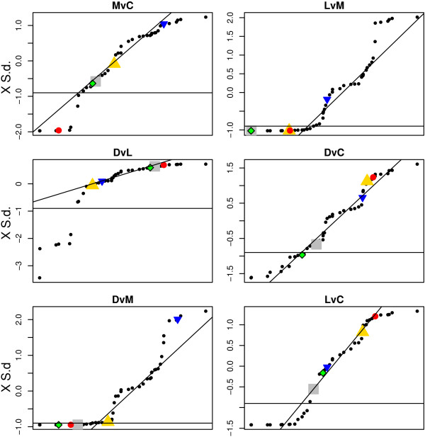
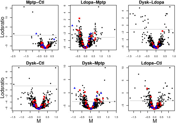
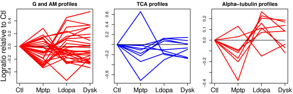
Using 2D-DIGE we analysed the protein content of the striatum from 6 control and 21 MPTP-treated monkeys, with or without de novo or long-term L-DOPA administration. There was an intensity and spatial bias in the data of all the gels examined in this study. Only two of the eight normalization methods evaluated ('2D loess+scale' and 'SC-2D+quantile') successfully removed both the intensity and spatial bias. In 'SC-2D+quantile' we extended the commonly used loess normalization method against dye bias in two-channel microarray systems to suit systems with three or more channels.Further, by using the proposed method, Differential Expression in Predefined Proteins Sets (DEPPS), several sets of proteins associated with the priming effects of L-DOPA in the striatum in parkinsonian animals were identified. Three of these sets are proteins involved in energy metabolism and one set involved proteins which are part of the microtubule cytoskeleton.

CONCLUSION

Comparison of the different methods leads to a series of methodological recommendations for the normalization and the analysis of data, depending on the experimental design. Due to the nature of 2D-DIGE data we recommend that the p-values obtained in significance tests should be used as rankings only. Individual proteins may be interesting as such, but by studying sets of proteins the interpretation of the results are probably more accurate and biologically informative.

摘要

背景

二维差异凝胶电泳(2D-DIGE)是用于测量样本或条件之间蛋白质表达差异的强大工具。然而,为了消除凝胶内部和之间的系统变异性,数据必须进行归一化处理。在本研究中,我们检验了四种现有和四种新型归一化方法消除2D-DIGE产生的数据中系统偏差的能力。我们还提出了对现有方法的一种改进,其中统计框架确定一组蛋白质是否与感兴趣的预定义表型相关联。该方法应用于我们从帕金森病猕猴模型(食蟹猴)生成的数据。

结果

使用2D-DIGE,我们分析了6只对照猴和21只经MPTP处理的猴纹状体的蛋白质含量,这些猴接受或未接受从头或长期左旋多巴给药。在本研究中检测的所有凝胶数据中存在强度和空间偏差。评估的八种归一化方法中只有两种(“二维局部加权回归+缩放”和“SC-2D+分位数”)成功消除了强度和空间偏差。在“SC-2D+分位数”中,我们将常用于消除双通道微阵列系统中染料偏差的局部加权回归归一化方法扩展到适用于具有三个或更多通道的系统。此外,通过使用所提出的方法,即预定义蛋白质组中的差异表达(DEPPS),鉴定了与帕金森病动物纹状体中左旋多巴引发效应相关的几组蛋白质。其中三组是参与能量代谢的蛋白质,一组是参与微管细胞骨架的蛋白质。

结论

根据实验设计,不同方法的比较产生了一系列关于数据归一化和分析的方法学建议。由于2D-DIGE数据的性质,我们建议在显著性检验中获得的p值仅用作排名。单个蛋白质本身可能很有趣,但通过研究蛋白质组,结果的解释可能更准确且具有生物学信息性。

https://cdn.ncbi.nlm.nih.gov/pmc/blobs/1958/1635739/7f27f24a4d7d/1471-2105-7-475-11.jpg
https://cdn.ncbi.nlm.nih.gov/pmc/blobs/1958/1635739/7df972f30737/1471-2105-7-475-1.jpg
https://cdn.ncbi.nlm.nih.gov/pmc/blobs/1958/1635739/5bbca0166a70/1471-2105-7-475-2.jpg
https://cdn.ncbi.nlm.nih.gov/pmc/blobs/1958/1635739/4abd4f6c3957/1471-2105-7-475-3.jpg
https://cdn.ncbi.nlm.nih.gov/pmc/blobs/1958/1635739/40f5830099af/1471-2105-7-475-4.jpg
https://cdn.ncbi.nlm.nih.gov/pmc/blobs/1958/1635739/d0dfb41b6bcd/1471-2105-7-475-5.jpg
https://cdn.ncbi.nlm.nih.gov/pmc/blobs/1958/1635739/32159a225dfe/1471-2105-7-475-6.jpg
https://cdn.ncbi.nlm.nih.gov/pmc/blobs/1958/1635739/596b8923ec8f/1471-2105-7-475-7.jpg
https://cdn.ncbi.nlm.nih.gov/pmc/blobs/1958/1635739/25b51f178731/1471-2105-7-475-8.jpg
https://cdn.ncbi.nlm.nih.gov/pmc/blobs/1958/1635739/268cdc47d423/1471-2105-7-475-9.jpg
https://cdn.ncbi.nlm.nih.gov/pmc/blobs/1958/1635739/c7bbfefa3d67/1471-2105-7-475-10.jpg
https://cdn.ncbi.nlm.nih.gov/pmc/blobs/1958/1635739/6dc7786ce0a2/1471-2105-7-475-12.jpg
https://cdn.ncbi.nlm.nih.gov/pmc/blobs/1958/1635739/7f27f24a4d7d/1471-2105-7-475-11.jpg

相似文献

1
Normalization and expression changes in predefined sets of proteins using 2D gel electrophoresis: a proteomic study of L-DOPA induced dyskinesia in an animal model of Parkinson's disease using DIGE.使用二维凝胶电泳对预定义蛋白质组进行标准化及表达变化分析:一项利用差异凝胶电泳(DIGE)技术对帕金森病动物模型中左旋多巴诱导的异动症进行蛋白质组学研究。
BMC Bioinformatics. 2006 Oct 26;7:475. doi: 10.1186/1471-2105-7-475.
2
Striatal proteomic analysis suggests that first L-dopa dose equates to chronic exposure.纹状体蛋白质组学分析表明,首次左旋多巴剂量等同于长期暴露。
PLoS One. 2008 Feb 13;3(2):e1589. doi: 10.1371/journal.pone.0001589.
3
Brain α7 nicotinic acetylcholine receptors in MPTP-lesioned monkeys and parkinsonian patients.MPTP 损毁猴和帕金森病患者脑内 α7 型烟碱型乙酰胆碱受体。
Biochem Pharmacol. 2016 Jun 1;109:62-69. doi: 10.1016/j.bcp.2016.03.023. Epub 2016 Mar 30.
4
Effects of L-DOPA-therapy on dopamine D2 receptor mRNA expression in the striatum of MPTP-intoxicated parkinsonian monkeys.左旋多巴治疗对MPTP中毒帕金森病猴纹状体中多巴胺D2受体mRNA表达的影响。
Brain Res Mol Brain Res. 1996 Nov;42(1):149-55. doi: 10.1016/s0169-328x(96)00157-x.
5
Native DIGE proteomic analysis of mitochondria from substantia nigra and striatum during neuronal degeneration and its compensation in an animal model of early Parkinson's disease.在早期帕金森病动物模型中,对黑质和纹状体神经元变性及其代偿过程中线粒体进行的天然差异凝胶电泳蛋白质组学分析。
Arch Physiol Biochem. 2016 Dec;122(5):238-256. doi: 10.1080/13813455.2016.1197948. Epub 2016 Jul 28.
6
Chronic treatment with MPEP, an mGlu5 receptor antagonist, normalizes basal ganglia glutamate neurotransmission in L-DOPA-treated parkinsonian monkeys.慢性给予 MPEP(一种 mGlu5 受体拮抗剂)可使帕金森病猴接受左旋多巴治疗后的基底神经节谷氨酸能神经传递正常化。
Neuropharmacology. 2013 Oct;73:216-31. doi: 10.1016/j.neuropharm.2013.05.028. Epub 2013 Jun 10.
7
The effects of chronic levodopa treatment on pre- and postsynaptic markers of dopaminergic function in striatum of parkinsonian monkeys.慢性左旋多巴治疗对帕金森病猴纹状体中多巴胺能功能的突触前和突触后标志物的影响。
Mov Disord. 1997 Mar;12(2):148-58. doi: 10.1002/mds.870120204.
8
GDNF reverses priming for dyskinesia in MPTP-treated, L-DOPA-primed common marmosets.胶质细胞源性神经营养因子可逆转经1-甲基-4-苯基-1,2,3,6-四氢吡啶(MPTP)处理并用左旋多巴(L-DOPA)引发异动症的普通狨猴的异动症致敏状态。
Eur J Neurosci. 2001 Feb;13(3):597-608. doi: 10.1046/j.1460-9568.2001.01408.x.
9
Differential proteomic and genomic profiling of mouse striatal cell model of Huntington's disease and control; probable implications to the disease biology.亨廷顿舞蹈病小鼠纹状体细胞模型与对照的差异蛋白质组学和基因组分析;对疾病生物学的可能影响。
J Proteomics. 2016 Jan 30;132:155-66. doi: 10.1016/j.jprot.2015.11.007. Epub 2015 Nov 12.
10
Striatal Akt/GSK3 signaling pathway in the development of L-Dopa-induced dyskinesias in MPTP monkeys.纹状体 Akt/GSK3 信号通路在 MPTP 猴模型中左旋多巴诱导的运动障碍发展中的作用。
Prog Neuropsychopharmacol Biol Psychiatry. 2010 Apr 16;34(3):446-54. doi: 10.1016/j.pnpbp.2009.12.011. Epub 2009 Dec 16.

引用本文的文献

1
Anatomo-proteomic characterization of human basal ganglia: focus on striatum and globus pallidus.人类基底神经节的解剖蛋白质组学特征:聚焦于纹状体和苍白球。
Mol Brain. 2014 Nov 18;7:83. doi: 10.1186/s13041-014-0083-9.
2
Infection with dengue-2 virus alters proteins in naturally expectorated saliva of Aedes aegypti mosquitoes.登革热2型病毒感染会改变埃及伊蚊自然咳出的唾液中的蛋白质。
Parasit Vectors. 2014 May 30;7:252. doi: 10.1186/1756-3305-7-252.
3
Proteomic analysis of striatum from MPTP-treated marmosets (Callithrix jacchus) with L-DOPA-induced dyskinesia of differing severity.

本文引用的文献

1
Enhanced preproenkephalin-B-derived opioid transmission in striatum and subthalamic nucleus converges upon globus pallidus internalis in L-3,4-dihydroxyphenylalanine-induced dyskinesia.在L-3,4-二羟基苯丙氨酸诱导的异动症中,纹状体和丘脑底核中增强的前脑啡肽原B衍生的阿片样物质传递汇聚于苍白球内侧部。
Biol Psychiatry. 2007 Apr 1;61(7):836-44. doi: 10.1016/j.biopsych.2006.06.038. Epub 2006 Sep 1.
2
Linear models and empirical bayes methods for assessing differential expression in microarray experiments.用于评估微阵列实验中差异表达的线性模型和经验贝叶斯方法。
Stat Appl Genet Mol Biol. 2004;3:Article3. doi: 10.2202/1544-6115.1027. Epub 2004 Feb 12.
3
对经MPTP处理且患有不同严重程度左旋多巴诱导性运动障碍的狨猴(狨属)纹状体进行蛋白质组学分析。
J Mol Neurosci. 2014 Feb;52(2):302-12. doi: 10.1007/s12031-013-0145-y. Epub 2013 Oct 23.
4
Brain region specific mitophagy capacity could contribute to selective neuronal vulnerability in Parkinson's disease.脑区特异性线粒体自噬能力可能导致帕金森病中神经元的选择性易损性。
Proteome Sci. 2011 Sep 23;9:59. doi: 10.1186/1477-5956-9-59.
5
Impact of temperature dependent sampling procedures in proteomics and peptidomics--a characterization of the liver and pancreas post mortem degradome.温度依赖型采样程序对蛋白质组学和肽组学的影响——对死后肝脏和胰腺降解组的特征描述。
Mol Cell Proteomics. 2011 Mar;10(3):M900229MCP200. doi: 10.1074/mcp.M900229-MCP200. Epub 2010 Jan 28.
6
Integrative analysis of the heat shock response in Aspergillus fumigatus.整合分析烟曲霉的热休克反应。
BMC Genomics. 2010 Jan 15;11:32. doi: 10.1186/1471-2164-11-32.
7
Priming for l-dopa-induced dyskinesia in Parkinson's disease: a feature inherent to the treatment or the disease?帕金森病中左旋多巴诱导的异动症的启动:是治疗固有的特征还是疾病本身的特征?
Prog Neurobiol. 2009 Jan 12;87(1):1-9. doi: 10.1016/j.pneurobio.2008.09.013. Epub 2008 Sep 30.
8
Striatal proteomic analysis suggests that first L-dopa dose equates to chronic exposure.纹状体蛋白质组学分析表明,首次左旋多巴剂量等同于长期暴露。
PLoS One. 2008 Feb 13;3(2):e1589. doi: 10.1371/journal.pone.0001589.
Proteomics-driven progress in neurodegeneration research.
蛋白质组学推动神经退行性疾病研究进展。
Electrophoresis. 2006 Apr;27(8):1556-73. doi: 10.1002/elps.200500738.
4
Proteomic evaluation of neonatal exposure to 2,2 ,4,4 ,5-pentabromodiphenyl ether.新生儿暴露于2,2',4,4',5-五溴二苯醚的蛋白质组学评估
Environ Health Perspect. 2006 Feb;114(2):254-9. doi: 10.1289/ehp.8419.
5
Effects of post-electrophoretic analysis on variance in gel-based proteomics.凝胶蛋白质组学中电泳后分析对方差的影响。
Expert Rev Proteomics. 2006 Feb;3(1):129-42. doi: 10.1586/14789450.3.1.129.
6
The Gene Ontology (GO) project in 2006.2006年的基因本体论(GO)项目。
Nucleic Acids Res. 2006 Jan 1;34(Database issue):D322-6. doi: 10.1093/nar/gkj021.
7
Software-induced variance in two-dimensional gel electrophoresis image analysis.二维凝胶电泳图像分析中的软件诱导变异
Electrophoresis. 2005 Dec;26(23):4508-20. doi: 10.1002/elps.200500253.
8
Impact of replicate types on proteomic expression analysis.重复类型对蛋白质组表达分析的影响。
J Proteome Res. 2005 Sep-Oct;4(5):1867-71. doi: 10.1021/pr050084g.
9
Modeling biological variability in 2-D gel proteomic carcinogenesis experiments.二维凝胶蛋白质组学致癌实验中的生物变异性建模
J Proteome Res. 2005 Sep-Oct;4(5):1619-27. doi: 10.1021/pr0501261.
10
Gene set enrichment analysis: a knowledge-based approach for interpreting genome-wide expression profiles.基因集富集分析:一种基于知识的方法用于解读全基因组表达谱。
Proc Natl Acad Sci U S A. 2005 Oct 25;102(43):15545-50. doi: 10.1073/pnas.0506580102. Epub 2005 Sep 30.