Defrate Louis E, Nha Kyung Wook, Papannagari Ramprasad, Moses Jeremy M, Gill Thomas J, Li Guoan
Bioengineering Laboratory, Massachusetts General Hospital and Harvard Medical School, 55 Fruit St, 1215 GRJ, Boston, MA 02114, USA.
J Biomech. 2007;40(8):1716-22. doi: 10.1016/j.jbiomech.2006.08.009. Epub 2006 Oct 27.
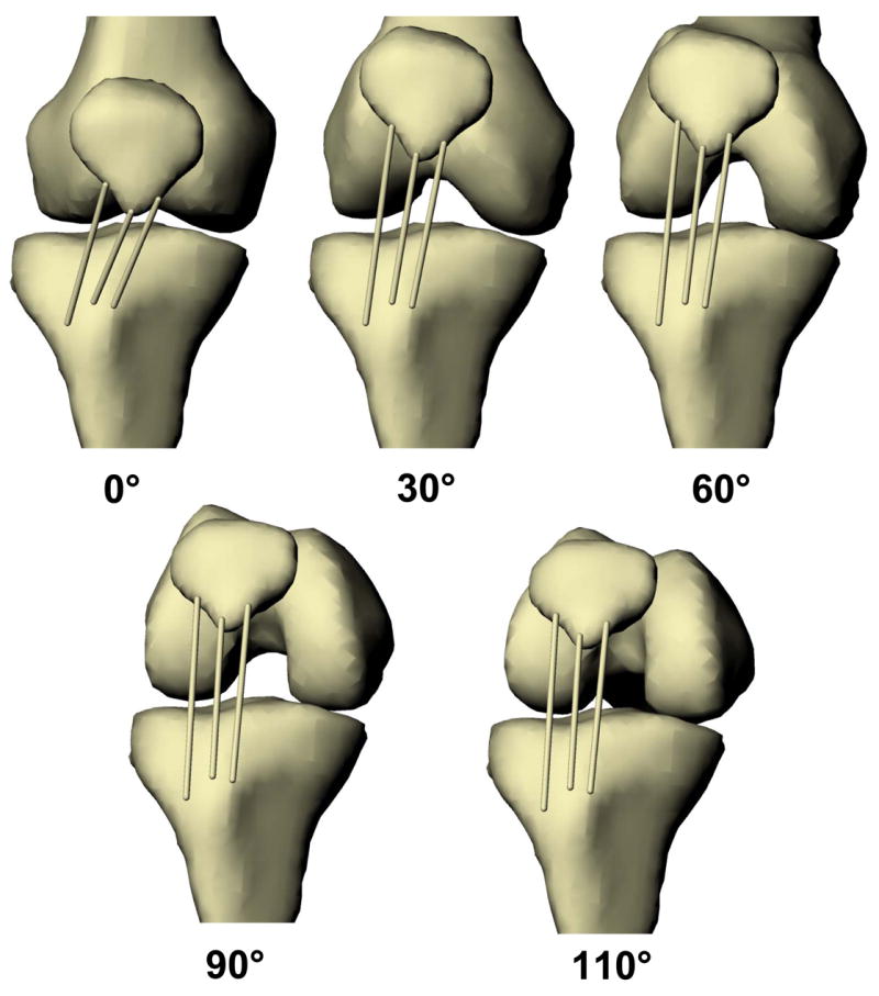
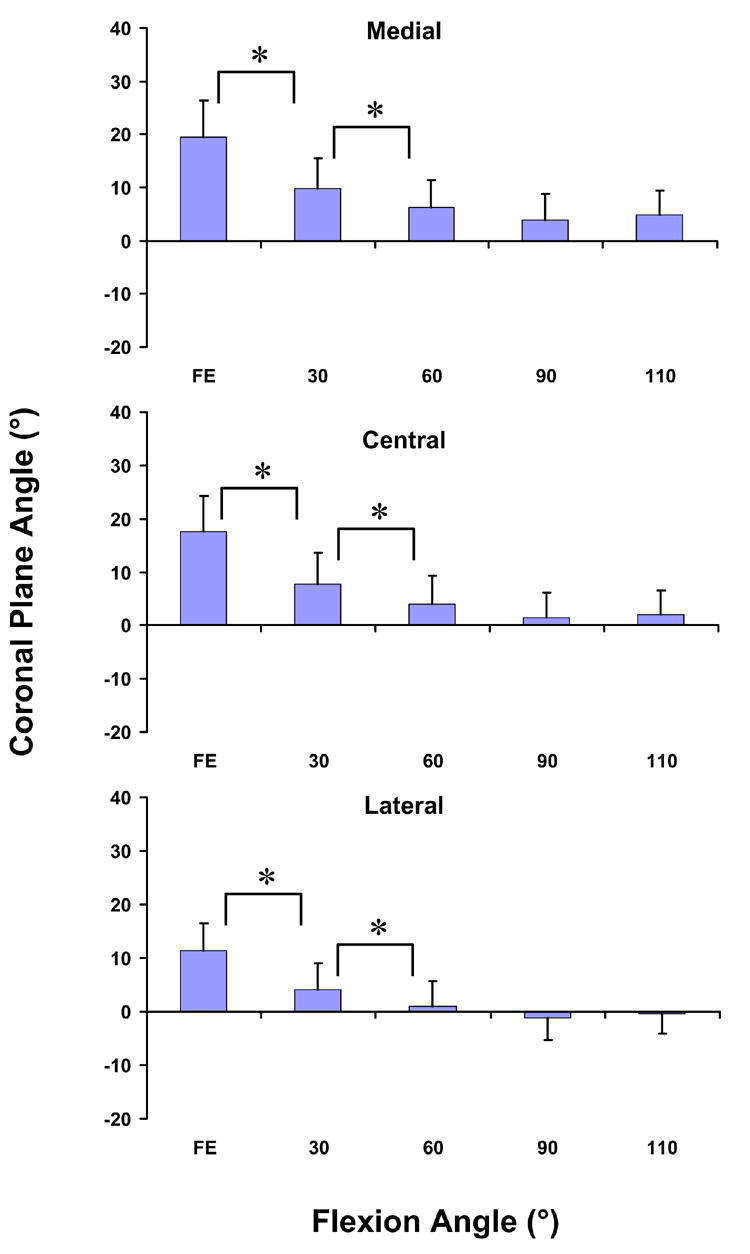
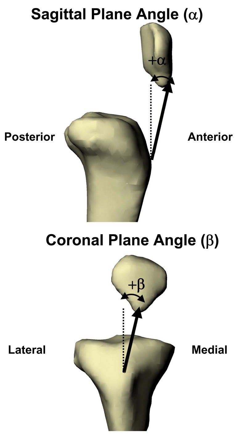
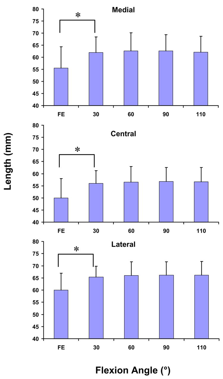
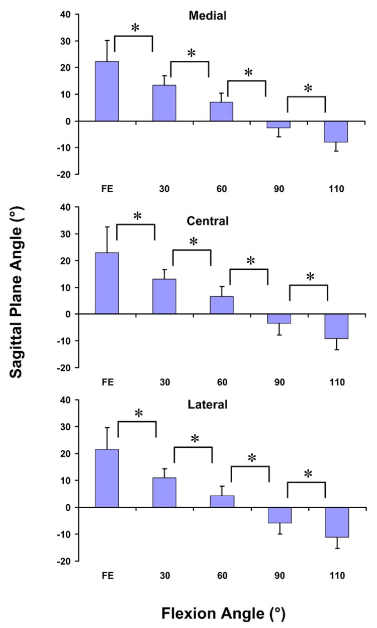
Few studies have investigated the function of the patellar tendon in-vivo. This study quantified the three-dimensional (3D) kinematics of the patellar tendon during weight-bearing flexion. Eleven subjects were imaged using magnetic resonance (MR). Sagittal plane images were outlined to create a 3D model of the patella, tibia, and femur and included the attachment sites of the patellar tendon. Each attachment site was divided into central, medial, and lateral thirds. Next, the subjects were imaged using fluoroscopy from two orthogonal directions while performing a single-leg lunge. The models and fluoroscopic images were used to reproduce the motion of the patella, tibia, and femur. The apparent elongation, sagittal plane angle, and coronal plane angle of each third of the patellar tendon were measured from the relative motion of the attachment sites. All three portions of the patellar tendon deformed similarly with flexion. The length of the patellar tendon significantly from full extension to 30 degrees . From 30 degrees -110 degrees , no significant change in the length of the patellar tendon was observed. The patellar tendon was oriented anteriorly at flexion angles less than 60 degrees and posteriorly thereafter. From full extension to 60 degrees , the medial orientation of the patellar tendon decreased significantly with flexion. These data may have important implications for anterior cruciate ligament reconstruction using patellar tendon autografts and for the design of rehabilitation regimens for patients of patellar tendon repair.
很少有研究在体内研究髌腱的功能。本研究量化了负重屈膝过程中髌腱的三维(3D)运动学。使用磁共振(MR)对11名受试者进行成像。勾勒矢状面图像以创建髌骨、胫骨和股骨的3D模型,并包括髌腱的附着部位。每个附着部位分为中央、内侧和外侧三分之一。接下来,在受试者进行单腿弓步时,从两个正交方向使用荧光透视成像。使用模型和荧光透视图像来重现髌骨、胫骨和股骨的运动。根据附着部位的相对运动测量髌腱各三分之一的表观伸长、矢状面角度和冠状面角度。髌腱的所有三个部分在屈膝时变形相似。从完全伸展到30度,髌腱长度显著变化。从30度到110度,未观察到髌腱长度有显著变化。屈膝角度小于60度时,髌腱向前定向,此后向后定向。从完全伸展到60度,髌腱的内侧定向随屈膝显著降低。这些数据可能对使用髌腱自体移植物进行前交叉韧带重建以及髌腱修复患者康复方案的设计具有重要意义。