Chen M C, Lee A T, Soll A H
Center for Ulcer Research and Education, Veterans Administration Wadsworth Hospital Center, Los Angeles, California 90073.
J Clin Invest. 1991 May;87(5):1716-23. doi: 10.1172/JCI115189.
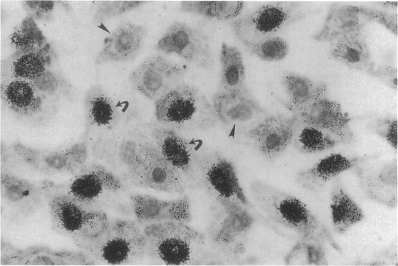
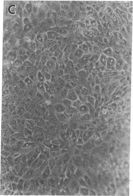
We report methods allowing the culture of rapidly dividing gastric epithelial cells to investigate the regulation of mucosal cell replication. Cells from canine fundic mucosa were dispersed by enzyme treatment, enriched by filtration and elutriation, and cultured on collagen gel in DMEM/F12 medium. After 48 h, greater than 95% of the cells displayed immunoreactivity with antibody to cytokeratin, an epithelial marker. The cells formed confluent monolayers by 72 h with a transmembrane resistance of 1,600 ohm.cm2 when mounted in a Ussing chamber indicating retention of epithelial cell characteristics. Calf serum (0.1-2%) produced a dose-dependent mitogenic effect evident by increases in [3H]-thymidine incorporation into acid-precipitated material and in cell number. After an 18-24-h incubation with [3H]-thymidine, approximately 55% of the cells cultured in 2% serum showed evidence of DNA synthesis by autoradiography and all of the replicating cells were cytokeratin positive. Using comparable culture conditions, a similar proportion of cells incubated for 18-24 h with bromodeoxyuridine displayed nuclear anti-bromodeoxyuridine immunoreactivity, thus indicating that over half of the cells in these cultures synthesized DNA during this period. As with serum, epidermal growth factor and transforming growth factor alpha (TGF alpha) (10 pM to 1 nM), insulin (10 nM to 1 microM) and insulinlike growth factor-I (IGF-I, 1-100 nM) increased [3H]-thymidine uptake. The greater potency of IGF-I, compared to insulin, suggests the presence of IGF-I receptors. We conclude that this culture preparation is composed of fundic mucosal epithelial cells and contains a predominance of dividing epithelial cells. EGF/TGF alpha and IGF-I are potential factors directly regulating proliferation of fundic mucosal cells.
我们报告了一些方法,可用于培养快速分裂的胃上皮细胞,以研究黏膜细胞复制的调控机制。犬胃底黏膜细胞经酶处理后分散,通过过滤和淘洗进行富集,然后在DMEM/F12培养基中的胶原凝胶上培养。48小时后,超过95%的细胞与上皮标志物细胞角蛋白抗体呈现免疫反应性。72小时时细胞形成汇合单层,安装在尤斯灌流小室中时跨膜电阻为1600欧姆·平方厘米,表明保留了上皮细胞特征。小牛血清(0.1 - 2%)产生剂量依赖性促有丝分裂作用,表现为[³H] - 胸腺嘧啶核苷掺入酸沉淀物质以及细胞数量增加。用[³H] - 胸腺嘧啶核苷孵育18 - 24小时后,在2%血清中培养的细胞约55%通过放射自显影显示有DNA合成证据,且所有复制细胞均为细胞角蛋白阳性。使用类似培养条件,用溴脱氧尿苷孵育18 - 24小时的细胞有相似比例显示核抗溴脱氧尿苷免疫反应性,这表明在此期间这些培养物中超过一半的细胞合成了DNA。与血清一样,表皮生长因子和转化生长因子α(TGFα)(10 pM至1 nM)、胰岛素(10 nM至1 μM)和胰岛素样生长因子 - I(IGF - I,1 - 100 nM)增加了[³H] - 胸腺嘧啶核苷摄取。与胰岛素相比,IGF - I效力更强,提示存在IGF - I受体。我们得出结论,这种培养制剂由胃底黏膜上皮细胞组成,且含有大量正在分裂的上皮细胞。表皮生长因子/转化生长因子α和胰岛素样生长因子 - I是直接调节胃底黏膜细胞增殖的潜在因子。