de Abreu Marilda Aparecida Milanez Morgado, Michalany Nilceo Schwery, Weckx Luc Louis Maurice, Neto Pimentel Dalva Regina, Hirata Cleonice Hitomi Watashi, de Avelar Alchorne Maurício Mota
Department of Pathology, EPM, UNIFESP, Federal University of São Paulo, Paulista School of Medicine, Brazil.
Braz J Otorhinolaryngol. 2006 May-Jun;72(3):312-6. doi: 10.1016/s1808-8694(15)30962-9.
Multibacillary leprosy may involve the oral mucosa, with or without apparent lesions. There are few studies that deal with this issue in the era of multidrug therapy.
To assess the frequency of oral mucosa involvement in multibacillary leprosy patients.
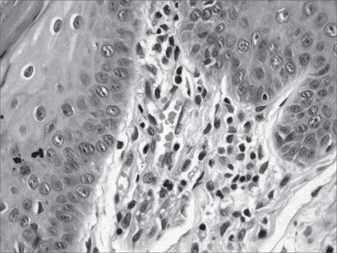
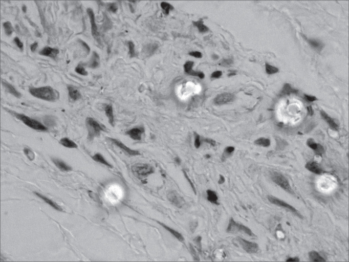
A transversal study with twenty non-treated multibacillary leprosy patients. The patients were treated in Dracena, São Paulo, between 2000 and 2002. Clinical examination of the oral mucosa was carried out. All patients were submitted to jugal mucosa, soft palate and tongue biopsies, in altered or in pre-established sites. The cross-sections were stained by techniques of hematoxilin-eosin and Ziehl-Neelsen. Granuloma and alcohol-acid-resistant bacilli findings determined the specific histopathological involvement.
The study involved 19 patients with an average of 2.5 years of disease progression. Specific histopathological involvement occurred in the tongue and soft palate of one lepromatous patient with an apparently normal oral mucosa.
(1) Clinical alterations in the oral mucosa does not imply disease involvement, it is necessary to have histopathological confirmation. (2) Apparent specific clinical alterations are rare. (3) The clinically normal oral mucosa can show specific histopathological involvement.
多菌型麻风可能累及口腔黏膜,伴或不伴有明显病变。在多药联合治疗时代,针对此问题的研究较少。
评估多菌型麻风患者口腔黏膜受累的频率。
对20例未经治疗的多菌型麻风患者进行横断面研究。这些患者于2000年至2002年期间在圣保罗州德拉塞纳接受治疗。对口腔黏膜进行临床检查。所有患者均在病变部位或预先设定的部位进行颊黏膜、软腭和舌活检。切片采用苏木精-伊红染色和齐-尼氏染色技术。肉芽肿和抗酸杆菌的发现确定了具体的组织病理学受累情况。
该研究纳入了19例患者,疾病进展平均时间为2.5年。一名口腔黏膜外观正常的瘤型麻风患者的舌部和软腭出现了特异性组织病理学受累情况。
(1)口腔黏膜的临床改变并不意味着疾病累及,必须进行组织病理学确诊。(2)明显的特异性临床改变很少见。(3)临床上正常的口腔黏膜可能出现特异性组织病理学受累情况。