• 文献检索
  • 文档翻译
  • 深度研究
  • 学术资讯
  • Suppr Zotero 插件Zotero 插件
  • 邀请有礼
  • 套餐&价格
  • 历史记录
应用&插件
Suppr Zotero 插件Zotero 插件浏览器插件Mac 客户端Windows 客户端微信小程序
定价
高级版会员购买积分包购买API积分包
服务
文献检索文档翻译深度研究API 文档MCP 服务
关于我们
关于 Suppr公司介绍联系我们用户协议隐私条款
关注我们

Suppr 超能文献

核心技术专利:CN118964589B侵权必究
粤ICP备2023148730 号-1Suppr @ 2026

文献检索

告别复杂PubMed语法,用中文像聊天一样搜索,搜遍4000万医学文献。AI智能推荐,让科研检索更轻松。

立即免费搜索

文件翻译

保留排版,准确专业,支持PDF/Word/PPT等文件格式,支持 12+语言互译。

免费翻译文档

深度研究

AI帮你快速写综述,25分钟生成高质量综述,智能提取关键信息,辅助科研写作。

立即免费体验

巨噬细胞浸润导致 Pendred 综合征小鼠模型中血管纹的退变。

Macrophage invasion contributes to degeneration of stria vascularis in Pendred syndrome mouse model.

作者信息

Jabba Sairam V, Oelke Alisha, Singh Ruchira, Maganti Rajanikanth J, Fleming Sherry, Wall Susan M, Everett Lorraine A, Green Eric D, Wangemann Philine

机构信息

Anatomy & Physiology Department, Kansas State University, Manhattan, KS 66506, USA.

出版信息

BMC Med. 2006 Dec 22;4:37. doi: 10.1186/1741-7015-4-37.

DOI:10.1186/1741-7015-4-37
PMID:17187680
原文链接:https://pmc.ncbi.nlm.nih.gov/articles/PMC1796619/
Abstract

BACKGROUND

Pendred syndrome, an autosomal-recessive disorder characterized by deafness and goiter, is caused by a mutation of SLC26A4, which codes for the anion exchanger pendrin. We investigated the relationship between pendrin expression and deafness using mice that have (Slc26a4+/+ or Slc26a4+/-) or lack (Slc26a4-/-) a complete Slc26a4 gene. Previously, we reported that stria vascularis of adult Slc26a4-/- mice is hyperpigmented and that marginal cells appear disorganized. Here we determine the time course of hyperpigmentation and marginal cell disorganization, and test the hypothesis that inflammation contributes to this tissue degeneration.

METHODS

Slc26a4-/- and age-matched control (Slc26a4+/+ or Slc26a4+/-) mice were studied at four postnatal (P) developmental stages: before and after the age that marks the onset of hearing (P10 and P15, respectively), after weaning (P28-41) and adult (P74-170). Degeneration and hyperpigmentation stria vascularis was evaluated by confocal microscopy. Gene expression in stria vascularis was analyzed by microarray and quantitative RT-PCR. In addition, the expression of a select group of genes was quantified in spiral ligament, spleen and liver to evaluate whether expression changes seen in stria vascularis are specific for stria vascularis or systemic in nature.

RESULTS

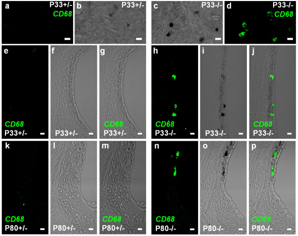
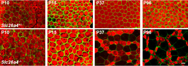
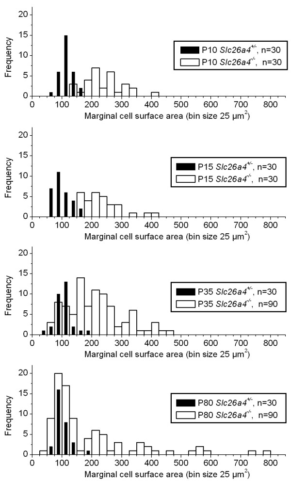
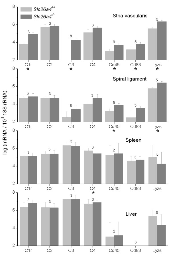
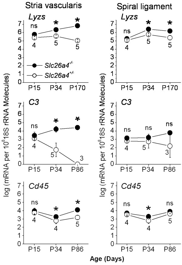
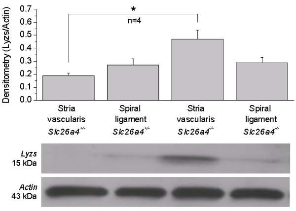
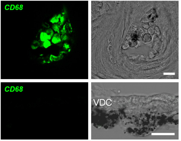
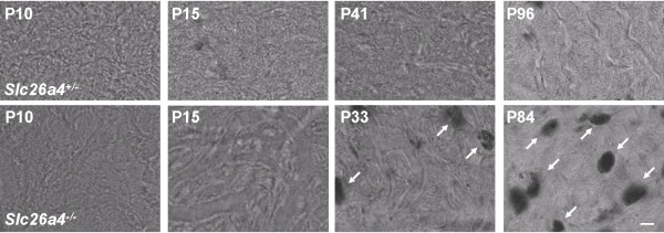
Degeneration of stria vascularis defined as hyperpigmentation and marginal cells disorganization was not seen at P10 or P15, but occurred after weaning and was associated with staining for CD68, a marker for macrophages. Marginal cells in Slc26a4-/-, however, had a larger apical surface area at P10 and P15. No difference in the expression of Lyzs, C3 and Cd45 was found in stria vascularis of P15 Slc26a4+/- and Slc26a4-/- mice. However, differences in expression were found after weaning and in adult mice. No difference in the expression of markers for acute inflammation, including Il1a, Il6, Il12a, Nos2 and Nos3 were found at P15, after weaning or in adults. The expression of macrophage markers including Ptprc (= Cd45), Cd68, Cd83, Lyzs, Lgals3 (= Mac2 antigen), Msr2, Cathepsins B, S, and K (Ctsb, Ctss, Ctsk) and complement components C1r, C3 and C4 was significantly increased in stria vascularis of adult Slc26a4-/- mice compared to Slc26a4+/+ mice. Expression of macrophage markers Cd45 and Cd84 and complement components C1r and C3 was increased in stria vascularis but not in spiral ligament, liver or spleen of Slc26a4-/- compared to Slc26a4+/- mice. The expression of Lyzs was increased in stria vascularis and spiral ligament but not in liver or spleen.

CONCLUSION

The data demonstrate that hyperpigmentation of stria vascularis and marginal cell reorganization in Slc26a4-/- mice occur after weaning, coinciding with an invasion of macrophages. The data suggest that macrophage invasion contributes to tissue degeneration in stria vascularis, and that macrophage invasion is restricted to stria vascularis and is not systemic in nature. The delayed onset of degeneration of stria vascularis suggests that a window of opportunity exists to restore/preserve hearing in mice and therefore possibly in humans suffering from Pendred syndrome.

摘要

背景

彭德莱德综合征是一种常染色体隐性疾病,其特征为耳聋和甲状腺肿,由编码阴离子交换蛋白pendrin的SLC26A4基因突变引起。我们利用具有完整Slc26a4基因(Slc26a4+/+或Slc26a4+/-)或缺乏该基因(Slc26a4-/-)的小鼠,研究了pendrin表达与耳聋之间的关系。此前,我们报道成年Slc26a4-/-小鼠的血管纹色素沉着过度,边缘细胞出现紊乱。在此,我们确定色素沉着过度和边缘细胞紊乱的时间进程,并检验炎症导致这种组织退化的假说。

方法

在四个出生后(P)发育阶段研究Slc26a4-/-小鼠和年龄匹配的对照小鼠(Slc26a4+/+或Slc26a4+/-):听力开始之前和之后(分别为P10和P15)、断奶后(P28 - 41)以及成年期(P74 - 170)。通过共聚焦显微镜评估血管纹的退化和色素沉着过度情况。通过微阵列和定量RT-PCR分析血管纹中的基因表达。此外,对螺旋韧带、脾脏和肝脏中一组选定基因的表达进行定量,以评估在血管纹中观察到的表达变化是血管纹特有的还是全身性的。

结果

在P10或P15时未观察到定义为色素沉着过度和边缘细胞紊乱的血管纹退化,但在断奶后出现,且与巨噬细胞标志物CD68的染色相关。然而,在P10和P15时,Slc26a4-/-小鼠的边缘细胞顶端表面积更大。在P15的Slc26a4+/-和Slc26a4-/-小鼠的血管纹中,未发现Lyzs、C3和Cd45表达有差异。然而,在断奶后和成年小鼠中发现了表达差异。在P15、断奶后或成年期,未发现包括Il1a、Il6、Il12a、Nos2和Nos3在内的急性炎症标志物表达有差异。与Slc26a4+/+小鼠相比,成年Slc26a4-/-小鼠血管纹中巨噬细胞标志物包括Ptprc(= Cd45)、Cd68、Cd83、Lyzs、Lgals3(= Mac2抗原)、Msr2、组织蛋白酶B、S和K(Ctsb、Ctss、Ctsk)以及补体成分C1r、C3和C4的表达显著增加。与Slc26a4+/-小鼠相比,Slc26a4-/-小鼠血管纹中巨噬细胞标志物Cd45和Cd84以及补体成分C1r和C3的表达增加,但在螺旋韧带、肝脏或脾脏中未增加。Lyzs的表达在血管纹和螺旋韧带中增加,但在肝脏或脾脏中未增加。

结论

数据表明,Slc26a4-/-小鼠血管纹色素沉着过度和边缘细胞重组在断奶后出现,与巨噬细胞浸润同时发生。数据提示巨噬细胞浸润导致血管纹组织退化,且巨噬细胞浸润仅限于血管纹,并非全身性的。血管纹退化的延迟发生表明存在恢复/保留小鼠听力的机会窗口,因此对于患有彭德莱德综合征的人类可能也存在这样的机会。

https://cdn.ncbi.nlm.nih.gov/pmc/blobs/3306/1796619/953bb3024e77/1741-7015-4-37-8.jpg
https://cdn.ncbi.nlm.nih.gov/pmc/blobs/3306/1796619/b2b1822d22fc/1741-7015-4-37-1.jpg
https://cdn.ncbi.nlm.nih.gov/pmc/blobs/3306/1796619/380595518c61/1741-7015-4-37-2.jpg
https://cdn.ncbi.nlm.nih.gov/pmc/blobs/3306/1796619/e91240bc92f5/1741-7015-4-37-3.jpg
https://cdn.ncbi.nlm.nih.gov/pmc/blobs/3306/1796619/78621a704209/1741-7015-4-37-4.jpg
https://cdn.ncbi.nlm.nih.gov/pmc/blobs/3306/1796619/47117d486571/1741-7015-4-37-5.jpg
https://cdn.ncbi.nlm.nih.gov/pmc/blobs/3306/1796619/89035478b99b/1741-7015-4-37-6.jpg
https://cdn.ncbi.nlm.nih.gov/pmc/blobs/3306/1796619/962fcaf922dd/1741-7015-4-37-7.jpg
https://cdn.ncbi.nlm.nih.gov/pmc/blobs/3306/1796619/953bb3024e77/1741-7015-4-37-8.jpg

相似文献

1
Macrophage invasion contributes to degeneration of stria vascularis in Pendred syndrome mouse model.巨噬细胞浸润导致 Pendred 综合征小鼠模型中血管纹的退变。
BMC Med. 2006 Dec 22;4:37. doi: 10.1186/1741-7015-4-37.
2
Free radical stress-mediated loss of Kcnj10 protein expression in stria vascularis contributes to deafness in Pendred syndrome mouse model.血管纹中自由基应激介导的Kcnj10蛋白表达缺失导致彭德莱综合征小鼠模型耳聋。
Am J Physiol Renal Physiol. 2008 Jan;294(1):F139-48. doi: 10.1152/ajprenal.00433.2007. Epub 2007 Oct 24.
3
Loss of cochlear HCO3- secretion causes deafness via endolymphatic acidification and inhibition of Ca2+ reabsorption in a Pendred syndrome mouse model.在一个 Pendred 综合征小鼠模型中,耳蜗 HCO3- 分泌的丧失通过内淋巴酸化和抑制 Ca2+ 重吸收导致耳聋。
Am J Physiol Renal Physiol. 2007 May;292(5):F1345-53. doi: 10.1152/ajprenal.00487.2006. Epub 2007 Feb 13.
4
Loss of KCNJ10 protein expression abolishes endocochlear potential and causes deafness in Pendred syndrome mouse model.KCNJ10蛋白表达缺失消除了内淋巴电位并导致 Pendred 综合征小鼠模型耳聋。
BMC Med. 2004 Aug 20;2:30. doi: 10.1186/1741-7015-2-30.
5
Progressive irreversible hearing loss is caused by stria vascularis degeneration in an Slc26a4-insufficient mouse model of large vestibular aqueduct syndrome.在大前庭导水管综合征的Slc26a4基因功能不足小鼠模型中,进行性不可逆听力损失是由血管纹变性引起的。
Neuroscience. 2015 Dec 3;310:188-97. doi: 10.1016/j.neuroscience.2015.09.016. Epub 2015 Sep 9.
6
Epithelial cell stretching and luminal acidification lead to a retarded development of stria vascularis and deafness in mice lacking pendrin.上皮细胞拉伸和管腔酸化导致敲除 pendrin 的小鼠中 stria vascularis 发育迟缓及耳聋。
PLoS One. 2011 Mar 14;6(3):e17949. doi: 10.1371/journal.pone.0017949.
7
Single-cell RNA-sequencing of stria vascularis cells in the adult Slc26a4 mouse.成年 Slc26a4 小鼠血管纹细胞的单细胞 RNA 测序。
BMC Med Genomics. 2023 Jun 15;16(1):133. doi: 10.1186/s12920-023-01549-0.
8
Slc26a4-insufficiency causes fluctuating hearing loss and stria vascularis dysfunction.Slc26a4 功能不全可导致波动性听力损失和血管纹功能障碍。
Neurobiol Dis. 2014 Jun;66:53-65. doi: 10.1016/j.nbd.2014.02.002. Epub 2014 Feb 19.
9
Deafness in LIMP2-deficient mice due to early loss of the potassium channel KCNQ1/KCNE1 in marginal cells of the stria vascularis.由于血管纹边缘细胞中钾通道KCNQ1/KCNE1的早期丧失,LIMP2缺陷小鼠出现耳聋。
J Physiol. 2006 Oct 1;576(Pt 1):73-86. doi: 10.1113/jphysiol.2006.116889. Epub 2006 Aug 10.
10
Transcriptomic Analysis Reveals an Altered Hcy Metabolism in the Stria Vascularis of the Pendred Syndrome Mouse Model.转录组分析揭示了 Pendred 综合征小鼠模型血管纹中 Hcy 代谢的改变。
Neural Plast. 2021 Apr 17;2021:5585394. doi: 10.1155/2021/5585394. eCollection 2021.

引用本文的文献

1
The Blood-Labyrinth Barrier: Non-Invasive Delivery Strategies for Inner Ear Drug Delivery.血迷路屏障:内耳药物递送的非侵入性策略
Pharmaceutics. 2025 Apr 7;17(4):482. doi: 10.3390/pharmaceutics17040482.
2
Early control of cochlear viral load limits cochlear inflammation and prevents virus-induced sensorineural hearing loss.早期控制耳蜗病毒载量可限制耳蜗炎症,并预防病毒诱导的感音神经性听力损失。
J Neuroinflammation. 2025 Mar 23;22(1):92. doi: 10.1186/s12974-025-03416-4.
3
The Stria Vascularis: Renewed Attention on a Key Player in Age-Related Hearing Loss.

本文引用的文献

1
Intercalated cell H+/OH- transporter expression is reduced in Slc26a4 null mice.闰细胞H⁺/OH⁻转运体的表达在Slc26a4基因敲除小鼠中降低。
Am J Physiol Renal Physiol. 2005 Dec;289(6):F1262-72. doi: 10.1152/ajprenal.00206.2005. Epub 2005 Sep 6.
2
Macrophages in inflammation.炎症中的巨噬细胞。
Curr Drug Targets Inflamm Allergy. 2005 Jun;4(3):281-6. doi: 10.2174/1568010054022024.
3
Mononuclear phagocytes migrate into the murine cochlea after acoustic trauma.声学创伤后,单核吞噬细胞迁移至小鼠耳蜗。
血管纹:关注年龄相关性听力损失关键因素的新视角。
Int J Mol Sci. 2024 May 15;25(10):5391. doi: 10.3390/ijms25105391.
4
Macrophage-related immune responses in inner ear: a potential therapeutic target for sensorineural hearing loss.内耳中与巨噬细胞相关的免疫反应:感音神经性听力损失的潜在治疗靶点。
Front Neurosci. 2024 Jan 11;17:1339134. doi: 10.3389/fnins.2023.1339134. eCollection 2023.
5
Effects of Therapeutic Hypothermia on Macrophages in Mouse Cochlea Explants.治疗性低温对小鼠耳蜗外植体中巨噬细胞的影响。
Int J Mol Sci. 2023 May 16;24(10):8850. doi: 10.3390/ijms24108850.
6
Single-cell transcriptomic atlas of mouse cochlear aging.小鼠耳蜗衰老的单细胞转录组图谱。
Protein Cell. 2023 Apr 13;14(3):180-201. doi: 10.1093/procel/pwac058.
7
Visualization of macrophage subsets in the development of the fetal human inner ear.胎儿人内耳发育中巨噬细胞亚群的可视化。
Front Immunol. 2022 Sep 9;13:965196. doi: 10.3389/fimmu.2022.965196. eCollection 2022.
8
Tissue-Resident Macrophages in the Stria Vascularis.血管纹中的组织驻留巨噬细胞
Front Neurol. 2022 Feb 3;13:818395. doi: 10.3389/fneur.2022.818395. eCollection 2022.
9
Role of the Stria Vascularis in the Pathogenesis of Sensorineural Hearing Loss: A Narrative Review.血管纹在感音神经性听力损失发病机制中的作用:一项叙述性综述。
Front Neurosci. 2021 Nov 19;15:774585. doi: 10.3389/fnins.2021.774585. eCollection 2021.
10
Macrophages Are Dispensable for Postnatal Pruning of the Cochlear Ribbon Synapses.巨噬细胞对于耳蜗带状突触的出生后修剪并非必需。
Front Cell Neurosci. 2021 Oct 21;15:736120. doi: 10.3389/fncel.2021.736120. eCollection 2021.
J Comp Neurol. 2005 Aug 22;489(2):180-94. doi: 10.1002/cne.20619.
4
Histological basis of the porcine femoral artery for vascular research.用于血管研究的猪股动脉的组织学基础。
Anat Histol Embryol. 2005 Apr;34(2):105-11. doi: 10.1111/j.1439-0264.2004.00580.x.
5
Innate immunity contributes to cochlear adaptive immune responses.先天免疫有助于耳蜗适应性免疫反应。
Audiol Neurootol. 2005 Jan-Feb;10(1):35-43. doi: 10.1159/000082306. Epub 2004 Nov 23.
6
Screening of SLC26A4 (PDS) gene in Pendred's syndrome: a large spectrum of mutations in France and phenotypic heterogeneity.佩德雷德综合征中SLC26A4(PDS)基因的筛查:法国的大量突变及表型异质性
Clin Genet. 2004 Oct;66(4):333-40. doi: 10.1111/j.1399-0004.2004.00296.x.
7
Loss of KCNJ10 protein expression abolishes endocochlear potential and causes deafness in Pendred syndrome mouse model.KCNJ10蛋白表达缺失消除了内淋巴电位并导致 Pendred 综合征小鼠模型耳聋。
BMC Med. 2004 Aug 20;2:30. doi: 10.1186/1741-7015-2-30.
8
Localization and functional studies of pendrin in the mouse inner ear provide insight about the etiology of deafness in pendred syndrome.小鼠内耳中 Pendrin 的定位与功能研究为 Pendred 综合征耳聋的病因提供了见解。
J Assoc Res Otolaryngol. 2003 Sep;4(3):394-404. doi: 10.1007/s10162-002-3052-4.
9
ASSOCIATION OF CONGENITAL DEAFNESS WITH GOITRE (PENDRED'S SYNDROME) A STUDY OF 207 FAMILIES.先天性耳聋与甲状腺肿的关联(彭德莱德综合征):对207个家庭的研究
Ann Hum Genet. 1965 Mar;28:201-49. doi: 10.1111/j.1469-1809.1964.tb00479.x.
10
Sugar transport by mammalian members of the SLC26 superfamily of anion-bicarbonate exchangers.SLC26 阴离子 - 碳酸氢根交换体超家族的哺乳动物成员介导的糖转运。
J Physiol. 2003 Aug 1;550(Pt 3):667-77. doi: 10.1113/jphysiol.2003.039321.