• 文献检索
  • 文档翻译
  • 深度研究
  • 学术资讯
  • Suppr Zotero 插件Zotero 插件
  • 邀请有礼
  • 套餐&价格
  • 历史记录
应用&插件
Suppr Zotero 插件Zotero 插件浏览器插件Mac 客户端Windows 客户端微信小程序
定价
高级版会员购买积分包购买API积分包
服务
文献检索文档翻译深度研究API 文档MCP 服务
关于我们
关于 Suppr公司介绍联系我们用户协议隐私条款
关注我们

Suppr 超能文献

核心技术专利:CN118964589B侵权必究
粤ICP备2023148730 号-1Suppr @ 2026

文献检索

告别复杂PubMed语法,用中文像聊天一样搜索,搜遍4000万医学文献。AI智能推荐,让科研检索更轻松。

立即免费搜索

文件翻译

保留排版,准确专业,支持PDF/Word/PPT等文件格式,支持 12+语言互译。

免费翻译文档

深度研究

AI帮你快速写综述,25分钟生成高质量综述,智能提取关键信息,辅助科研写作。

立即免费体验

多发性硬化症脑死后的扩散张量成像

Diffusion tensor imaging of post mortem multiple sclerosis brain.

作者信息

Schmierer Klaus, Wheeler-Kingshott Claudia A M, Boulby Phil A, Scaravilli Francesco, Altmann Daniel R, Barker Gareth J, Tofts Paul S, Miller David H

机构信息

Institute of Neurology, University College London, NMR Research Unit, Box 117, Queen Square, London WC1N 3BG, UK.

出版信息

Neuroimage. 2007 Apr 1;35(2):467-77. doi: 10.1016/j.neuroimage.2006.12.010. Epub 2006 Dec 16.

DOI:10.1016/j.neuroimage.2006.12.010
PMID:17258908
原文链接:https://pmc.ncbi.nlm.nih.gov/articles/PMC1892244/
Abstract

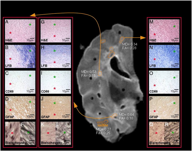
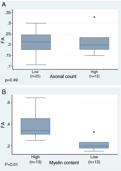
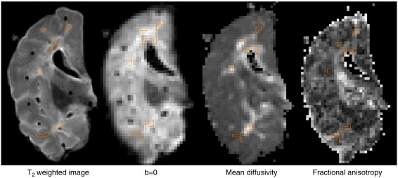
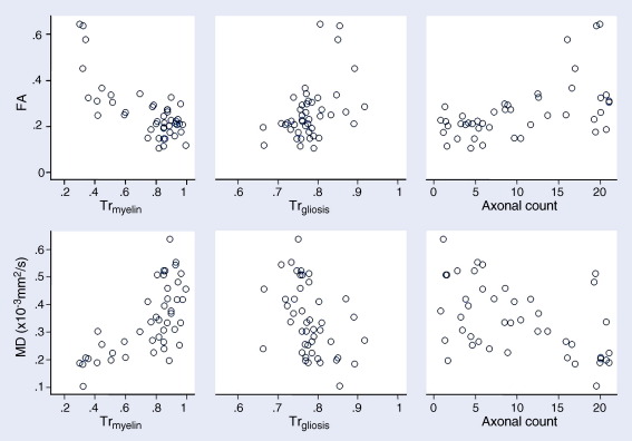
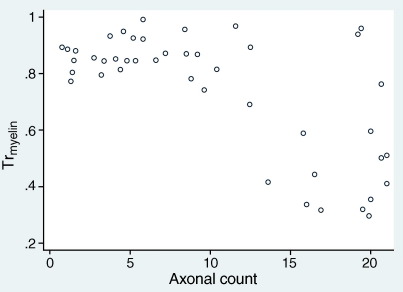
Magnetic resonance imaging (MRI) is being used to probe the central nervous system (CNS) of patients with multiple sclerosis (MS), a chronic demyelinating disease. Conventional T(2)-weighted MRI (cMRI) largely fails to predict the degree of patients' disability. This shortcoming may be due to poor specificity of cMRI for clinically relevant pathology. Diffusion tensor imaging (DTI) has shown promise to be more specific for MS pathology. In this study we investigated the association between histological indices of myelin content, axonal count and gliosis, and two measures of DTI (mean diffusivity [MD] and fractional anisotropy [FA]), in unfixed post mortem MS brain using a 1.5-T MR system. Both MD and FA were significantly lower in post mortem MS brain compared to published data acquired in vivo. However, the differences of MD and FA described in vivo between white matter lesions (WMLs) and normal-appearing white matter (NAWM) were retained in this study of post mortem brain: average MD in WMLs was 0.35x10(-3) mm(2)/s (SD, 0.09) versus 0.22 (0.04) in NAWM; FA was 0.22 (0.06) in WMLs versus 0.38 (0.13) in NAWM. Correlations were detected between myelin content (Tr(myelin)) and (i) FA (r=-0.79, p<0.001), (ii) MD (r=0.68, p<0.001), and (iii) axonal count (r=-0.81, p<0.001). Multiple regression suggested that these correlations largely explain the apparent association of axonal count with (i) FA (r=0.70, p<0.001) and (ii) MD (r=-0.66, p<0.001). In conclusion, this study suggests that FA and MD are affected by myelin content and - to a lesser degree - axonal count in post mortem MS brain.

摘要

磁共振成像(MRI)正被用于探测患有多发性硬化症(MS)的患者的中枢神经系统(CNS),多发性硬化症是一种慢性脱髓鞘疾病。传统的T(2)加权MRI(cMRI)在很大程度上无法预测患者的残疾程度。这一缺点可能是由于cMRI对临床相关病理的特异性较差。扩散张量成像(DTI)已显示出对MS病理更具特异性的前景。在本研究中,我们使用1.5-T MR系统,研究了未固定的死后MS脑内髓鞘含量、轴突计数和胶质增生的组织学指标与DTI的两项测量值(平均扩散率[MD]和分数各向异性[FA])之间的关联。与已发表的体内采集数据相比,死后MS脑内的MD和FA均显著降低。然而,在本死后脑研究中,保留了体内描述的白质病变(WMLs)和正常外观白质(NAWM)之间MD和FA的差异:WMLs中的平均MD为0.35×10(-3)mm(2)/s(标准差,0.09),而NAWM中的平均MD为0.22(0.04);WMLs中的FA为0.22(0.06),而NAWM中的FA为0.38(0.13)。在髓鞘含量(Tr(髓鞘))与(i)FA(r = -0.79,p < 0.001)、(ii)MD(r = 0.68,p < 0.001)和(iii)轴突计数(r = -0.81,p < 0.001)之间检测到相关性。多元回归表明,这些相关性在很大程度上解释了轴突计数与(i)FA(r = 0.70,p < 0.001)和(ii)MD(r = -0.66,p < 0.001)之间明显的关联。总之,本研究表明,在死后MS脑中,FA和MD受髓鞘含量影响,且在较小程度上受轴突计数影响。

https://cdn.ncbi.nlm.nih.gov/pmc/blobs/abc1/2148029/d8bc151b66e0/gr5.jpg
https://cdn.ncbi.nlm.nih.gov/pmc/blobs/abc1/2148029/acda004dd291/gr1.jpg
https://cdn.ncbi.nlm.nih.gov/pmc/blobs/abc1/2148029/4e6dd9fa5d84/gr2.jpg
https://cdn.ncbi.nlm.nih.gov/pmc/blobs/abc1/2148029/ed3ab17a3ad8/gr3.jpg
https://cdn.ncbi.nlm.nih.gov/pmc/blobs/abc1/2148029/07a366c9f2a2/gr4.jpg
https://cdn.ncbi.nlm.nih.gov/pmc/blobs/abc1/2148029/d8bc151b66e0/gr5.jpg

相似文献

1
Diffusion tensor imaging of post mortem multiple sclerosis brain.多发性硬化症脑死后的扩散张量成像
Neuroimage. 2007 Apr 1;35(2):467-77. doi: 10.1016/j.neuroimage.2006.12.010. Epub 2006 Dec 16.
2
Multiple sclerosis: hyperintense lesions in the brain on T1-weighted MR images assessed by diffusion tensor imaging.多发性硬化症:应用弥散张量成像评估 T1 加权 MR 图像上的脑高信号病变。
J Magn Reson Imaging. 2010 Apr;31(4):789-95. doi: 10.1002/jmri.22103.
3
Neurite orientation dispersion and density imaging in myelin oligodendrocyte glycoprotein antibody-associated disease and neuromyelitis optica spectrum disorders.髓鞘少突胶质细胞糖蛋白抗体相关疾病和视神经脊髓炎谱系障碍中的神经突方向离散度与密度成像
Mult Scler Relat Disord. 2025 Mar;95:106324. doi: 10.1016/j.msard.2025.106324. Epub 2025 Feb 8.
4
Quantitative magnetic resonance of postmortem multiple sclerosis brain before and after fixation.固定前后多发性硬化症死后大脑的定量磁共振成像
Magn Reson Med. 2008 Feb;59(2):268-77. doi: 10.1002/mrm.21487.
5
Diffusion tensor imaging applications in multiple sclerosis patients using 3T magnetic resonance: a preliminary study.3T 磁共振弥散张量成像在多发性硬化患者中的应用:初步研究。
Eur Radiol. 2012 May;22(5):990-7. doi: 10.1007/s00330-011-2342-9. Epub 2011 Dec 9.
6
In vivo assessment of cervical cord damage in MS patients: a longitudinal diffusion tensor MRI study.多发性硬化症患者颈髓损伤的体内评估:一项纵向扩散张量磁共振成像研究
Brain. 2007 Aug;130(Pt 8):2211-9. doi: 10.1093/brain/awm110. Epub 2007 May 29.
7
HIV-associated alterations in normal-appearing white matter: a voxel-wise diffusion tensor imaging study.人类免疫缺陷病毒相关的正常外观白质改变:一项基于体素的扩散张量成像研究
J Acquir Immune Defic Syndr. 2007 Dec 15;46(5):564-73. doi: 10.1097/qai.0b013e318159d807.
8
Quantitative magnetization transfer imaging in postmortem multiple sclerosis brain.尸检多发性硬化症脑的定量磁化传递成像
J Magn Reson Imaging. 2007 Jul;26(1):41-51. doi: 10.1002/jmri.20984.
9
Diffusion tensor magnetic resonance imaging in multiple sclerosis.多发性硬化症中的扩散张量磁共振成像
Neurology. 2001 Feb 13;56(3):304-11. doi: 10.1212/wnl.56.3.304.
10
Microstructural alterations in different types of lesions and their perilesional white matter in relapsing-remitting multiple sclerosis based on diffusion kurtosis imaging.基于扩散峰度成像的复发缓解型多发性硬化症不同类型病灶及其病灶周围白质的微观结构改变。
Mult Scler Relat Disord. 2023 Mar;71:104572. doi: 10.1016/j.msard.2023.104572. Epub 2023 Feb 19.

引用本文的文献

1
Leveling up: along-level diffusion tensor imaging in the spinal cord of multiple sclerosis patients.升级:多发性硬化症患者脊髓中的纵向扩散张量成像
Front Neuroimaging. 2025 Aug 11;4:1599966. doi: 10.3389/fnimg.2025.1599966. eCollection 2025.
2
Tract-specific analysis of diffusion MRI at 3T detects cervical spinal cord aberrations in multiple sclerosis.3T磁共振扩散加权成像的脊髓特异性分析可检测多发性硬化症患者的颈髓异常。
Imaging Neurosci (Camb). 2025 Jul 7;3. doi: 10.1162/IMAG.a.72. eCollection 2025.
3
Comparative effectiveness of teriflunomide and ocrelizumab on smoldering activity in multiple sclerosis: an observational study in the Swiss Multiple Sclerosis Cohort.

本文引用的文献

1
Magnetic resonance imaging of the common marmoset head.普通狨猴头部的磁共振成像
Altern Lab Anim. 1998 May-Jun;26(3):343-56.
2
Diffusion tensor imaging in multiple sclerosis: assessment of regional differences in the axial plane within normal-appearing cervical spinal cord.多发性硬化症中的扩散张量成像:对正常外观的颈髓轴平面内区域差异的评估
AJNR Am J Neuroradiol. 2006 Jun-Jul;27(6):1189-93.
3
Probabilistic diffusion tractography: a potential tool to assess the rate of disease progression in amyotrophic lateral sclerosis.概率性扩散张量成像:一种评估肌萎缩侧索硬化症疾病进展速率的潜在工具。
特立氟胺与奥瑞珠单抗对多发性硬化症隐匿活动的比较疗效:瑞士多发性硬化症队列的一项观察性研究
J Neurol. 2025 Jul 2;272(8):491. doi: 10.1007/s00415-025-13221-x.
4
Diffusion Tensor Imaging Findings in Cerebral Sensorimotor Areas in Patients After Spinal Cord Injury Correlate With Neurophysiological Deficits.脊髓损伤患者大脑感觉运动区的扩散张量成像结果与神经生理缺陷相关。
Neurorehabil Neural Repair. 2025 Jun 19;39(9):15459683251345434. doi: 10.1177/15459683251345434.
5
Layer-Dependent Effect of Aβ-Pathology on Cortical Microstructure With Ex Vivo Human Brain Diffusion MRI at 7 Tesla.7特斯拉下利用离体人脑扩散磁共振成像研究Aβ病理对皮质微结构的层依赖性影响
Hum Brain Mapp. 2025 May;46(7):e70222. doi: 10.1002/hbm.70222.
6
White Matter Tract Degeneration in Multiple Sclerosis Patients With Progression Independent of Relapse Activity.进展与复发活动无关的多发性硬化症患者的白质束变性
Neurol Neuroimmunol Neuroinflamm. 2025 May;12(3):e200388. doi: 10.1212/NXI.0000000000200388. Epub 2025 Apr 16.
7
Correlations between postmortem quantitative MRI parameters and demyelination, axonal loss and gliosis in multiple sclerosis: A systematic review and meta-analysis.多发性硬化症中死后定量MRI参数与脱髓鞘、轴突丢失和胶质细胞增生之间的相关性:一项系统评价和荟萃分析。
Brain Imaging Behav. 2025 Apr;19(2):323-335. doi: 10.1007/s11682-025-00971-5. Epub 2025 Jan 27.
8
Preservation of Bilateral Corticospinal Projections from Injured Hemisphere After Perinatal Stroke.围产期卒中后损伤半球双侧皮质脊髓投射的保留
Brain Sci. 2025 Jan 17;15(1):82. doi: 10.3390/brainsci15010082.
9
High-resolution diffusion magnetic resonance imaging and spatial-transcriptomic in developing mouse brain.高分辨率弥散磁共振成像和空间转录组学在发育中小鼠大脑中的应用。
Neuroimage. 2024 Aug 15;297:120734. doi: 10.1016/j.neuroimage.2024.120734. Epub 2024 Jul 20.
10
Advanced MRI Techniques: Diagnosis and Follow-Up of Multiple Sclerosis.先进的磁共振成像技术:多发性硬化症的诊断与随访
Diagnostics (Basel). 2024 May 28;14(11):1120. doi: 10.3390/diagnostics14111120.
Brain. 2006 Jul;129(Pt 7):1859-71. doi: 10.1093/brain/awl100. Epub 2006 May 3.
4
Noninvasive detection of cuprizone induced axonal damage and demyelination in the mouse corpus callosum.非侵入性检测小鼠胼胝体中铜螯合剂诱导的轴突损伤和脱髓鞘
Magn Reson Med. 2006 Feb;55(2):302-8. doi: 10.1002/mrm.20774.
5
Applications of diffusion tensor MR imaging in multiple sclerosis.扩散张量磁共振成像在多发性硬化症中的应用。
Ann N Y Acad Sci. 2005 Dec;1064:202-19. doi: 10.1196/annals.1340.039.
6
Diffusion MRI in multiple sclerosis.多发性硬化症中的扩散磁共振成像
Neurology. 2005 Nov 22;65(10):1526-32. doi: 10.1212/01.wnl.0000184471.83948.e0.
7
Detecting axon damage in spinal cord from a mouse model of multiple sclerosis.在多发性硬化症小鼠模型中检测脊髓轴突损伤。
Neurobiol Dis. 2006 Mar;21(3):626-32. doi: 10.1016/j.nbd.2005.09.009. Epub 2005 Nov 17.
8
Diagnostic criteria for multiple sclerosis: 2005 revisions to the "McDonald Criteria".多发性硬化症的诊断标准:对“麦克唐纳标准”的2005年修订版。
Ann Neurol. 2005 Dec;58(6):840-6. doi: 10.1002/ana.20703.
9
'Importance sampling' in MS: use of diffusion tensor tractography to quantify pathology related to specific impairment.多发性硬化症中的“重要性采样”:利用扩散张量纤维束成像来量化与特定损伤相关的病理学特征。
J Neurol Sci. 2005 Oct 15;237(1-2):13-9. doi: 10.1016/j.jns.2005.04.019.
10
Intracortical lesions in multiple sclerosis: improved detection with 3D double inversion-recovery MR imaging.多发性硬化症的皮质内病变:通过三维双反转恢复磁共振成像提高检测率。
Radiology. 2005 Jul;236(1):254-60. doi: 10.1148/radiol.2361040450.