Fotou Maria, Oikonomou Vassiliki, Zagouri Flora, Sergentanis Theodoros N, Nonni Afroditi, Athanassiadou Pauline, Drouveli Theodora, Atsouris Efstratios, Kotzia Evagelia, Zografos George C
Department of Cytology, Hippocratio Hospital, Athens, Greece.
World J Surg Oncol. 2007 Apr 3;5:40. doi: 10.1186/1477-7819-5-40.
To evaluate imprint cytology in the context of specimens with microcalcifications derived from Vacuum-Assisted Breast Biopsy (VABB).
A total of 93 women with microcalcifications BI-RADS 3 and 4 underwent VABB and imprint samples were examined. VABB was performed on Fischer's table using 11-gauge Mammotome vacuum probes. A mammogram of the cores after the procedure confirmed the excision of microcalcifications. For the application of imprint cytology, the cores with microcalcifications confirmed by mammogram were gently rolled against glass microscope slides and thus imprint smears were made. For rapid preliminary diagnosis Diff-Quick stain, modified Papanicolaou stain and May Grunwald Giemsa were used. Afterwards, the core was dipped into a CytoRich Red Collection fluid for a few seconds in order to obtain samples with the use of the specimen wash. After the completion of cytological procedures, the core was prepared for routine histological study. The pathologist was blind to the preliminary cytological results. The cytological and pathological diagnoses were comparatively evaluated.
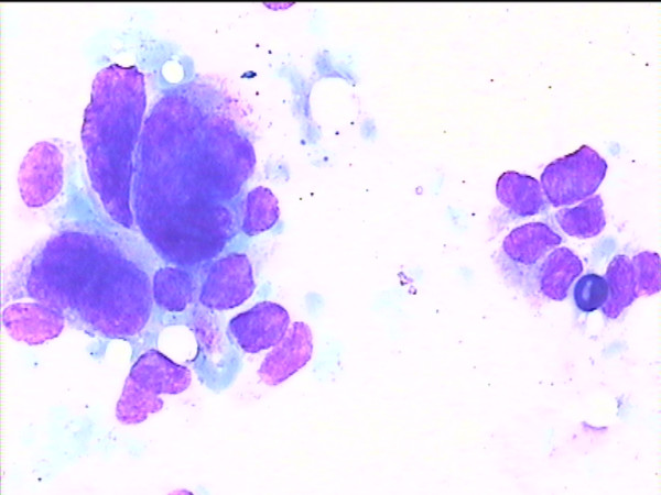
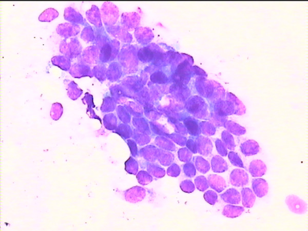
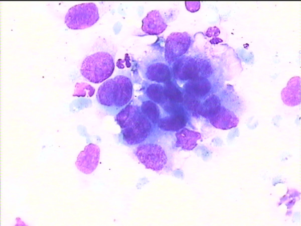
According to the pathological examination, 73 lesions were benign, 15 lesions were carcinomas (12 ductal carcinomas in situ, 3 invasive ductal carcinomas), and 5 lesions were precursor: 3 cases of atypical ductal hyperplasia (ADH) and 2 cases of lobular neoplasia (LN). The observed sensitivity and specificity of the cytological imprints for cancer were 100% (one-sided, 97.5% CI: 78.2%-100%). Only one case of ADH could be detected by imprint cytology. Neither of the two LN cases was detected by the imprints. The imprints were uninformative in 11 out of 93 cases (11.8%). There was no uninformative case among women with malignancy.
Imprint cytology provides a rapid, accurate preliminary diagnosis in a few minutes. This method might contribute to the diagnosis of early breast cancer and possibly attenuates patients' anxiety.
在真空辅助乳腺活检(VABB)获取的伴有微钙化的标本中评估印片细胞学。
93例BI-RADS 3级和4级微钙化女性接受了VABB检查,并对印片样本进行检测。VABB在Fischer手术台上使用11号麦默通真空探头进行。术后对标本条进行乳房X线摄影以确认微钙化灶已被切除。对于印片细胞学检查应用,将乳房X线摄影确认有微钙化的标本条在玻璃显微镜载玻片上轻轻滚动,从而制作印片涂片。采用Diff-Quick染色、改良巴氏染色和May Grunwald Giemsa染色进行快速初步诊断。之后,将标本条在CytoRich Red收集液中浸泡数秒,以便通过标本冲洗获取样本。细胞学检查完成后,将标本条制备用于常规组织学研究。病理科医生对初步细胞学结果不知情,并对细胞学和病理诊断进行比较评估。
根据病理检查,73个病变为良性病变,15个病变为癌(12例导管原位癌、3例浸润性导管癌),5个病变为癌前病变:3例非典型导管增生(ADH)和2例小叶瘤变(LN)。观察到印片细胞学检查对癌症的敏感性和特异性为100%(单侧,97.5%可信区间:78.2%-100%)。印片细胞学仅能检测出1例ADH。印片未检测出2例LN中的任何1例。93例中有11例(11.8%)印片结果无诊断价值。恶性病变女性中无印片结果无诊断价值的病例。
印片细胞学可在数分钟内提供快速、准确的初步诊断。该方法可能有助于早期乳腺癌的诊断,并可能减轻患者的焦虑。