Gutierrez Guillermo, Venbrux Anthony, Ignacio Elizabeth, Reiner Jonathan, Chawla Lakhmir, Desai Anish
Division of Pulmonary and Critical Care Medicine, Department of Medicine, The George Washington University Medical Center, Pennsylvania Avenue, NW Washington, District of Columbia 20037, USA.
Crit Care. 2007;11(2):R44. doi: 10.1186/cc5739.
Decreases in oxygen saturation (SO2) and lactate concentration [Lac] from superior vena cava (SVC) to pulmonary artery have been reported. These gradients (Delta SO2 and Delta[Lac]) are probably created by diluting SVC blood with blood of lower SO2 and [Lac]. We tested the hypothesis that Delta SO2 and Delta[Lac] result from mixing SVC and inferior vena cava (IVC) blood streams.
This was a prospective, sequential, observational study of hemodynamically stable individuals with pulmonary artery hypertension (n = 9) who were about to undergo right heart catheterization. Catheters were advanced under fluoroscopic guidance into the IVC, SVC, right atrium, right ventricle, and pulmonary artery. Samples were obtained at each site and analyzed for SO2, [Lac], and glucose concentration ([Glu]). Analysis of variance with Tukey HSD test was used to compare metabolite concentrations at each site.
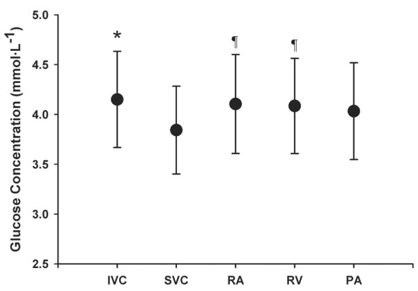
There were no differences in SO2 or [Lac] between IVC and SVC, both being greater than their respective pulmonary artery measurements (P < 0.01 for SO2 and P < 0.05 for [Lac]). SO2 and [Lac] in right atrium, right ventricle, and pulmonary artery were similar. Delta SO2 was 4.4 +/- 1.4% (mean +/- standard deviation) and Delta[Lac] was 0.16 +/- 0.11 mmol/l (both > 0; P < 0.001). Delta[Glu] was -0.19 +/- 0.31 mmol/l, which was not significantly different from zero, with SVC [Glu] being less than IVC [Glu].
Mixing of SVC with IVC blood does not account for the development of Delta SO2 and Delta[Lac] in hemodynamically stable individuals with pulmonary artery hypertension. An alternate mechanism is mixing with coronary sinus blood, implying that Delta SO2 and Delta[Lac] may reflect changes in coronary sinus SO2 and [Lac] in this patient population.
据报道,从上腔静脉(SVC)到肺动脉,氧饱和度(SO2)和乳酸浓度[Lac]会降低。这些梯度(ΔSO2和Δ[Lac])可能是由低SO2和[Lac]的血液稀释SVC血液所致。我们检验了这样一个假设,即ΔSO2和Δ[Lac]是由SVC和下腔静脉(IVC)血流混合导致的。
这是一项对9例即将接受右心导管检查的血流动力学稳定的肺动脉高压患者进行的前瞻性、连续性观察研究。在荧光镜引导下将导管推进至IVC、SVC、右心房、右心室和肺动脉。在每个部位采集样本并分析SO2、[Lac]和葡萄糖浓度([Glu])。采用方差分析和Tukey HSD检验比较各部位的代谢物浓度。
IVC和SVC之间的SO2或[Lac]无差异,两者均高于各自肺动脉的测量值(SO2,P<0.01;[Lac],P<0.05)。右心房、右心室和肺动脉中的SO2和[Lac]相似。ΔSO2为4.4±1.4%(均值±标准差),Δ[Lac]为0.16±0.11 mmol/l(两者均>0;P<0.001)。Δ[Glu]为-0.19±0.31 mmol/l,与零无显著差异,SVC的[Glu]低于IVC的[Glu]。
在血流动力学稳定的肺动脉高压患者中,SVC与IVC血液混合并不能解释ΔSO2和Δ[Lac]的产生。另一种机制是与冠状窦血液混合,这意味着ΔSO2和Δ[Lac]可能反映了该患者群体冠状窦SO2和[Lac]的变化。