Ando Seijitsu, Otani Hitomi, Yagi Yasuhiro, Kawai Kenzo, Araki Hiromasa, Fukuhara Shirou, Inagaki Chiyoko
Department of Pharmacology, Kansai Medical University, Fumizono-Cho, Moriguchi, Osaka, Japan.
Respir Res. 2007 Apr 16;8(1):31. doi: 10.1186/1465-9921-8-31.
Proteinase-activated receptors (PARs; PAR1-4) that can be activated by serine proteinases such as thrombin and neutrophil catepsin G are known to contribute to the pathogenesis of various pulmonary diseases including fibrosis. Among these PARs, especially PAR4, a newly identified subtype, is highly expressed in the lung. Here, we examined whether PAR4 stimulation plays a role in the formation of fibrotic response in the lung, through alveolar epithelial-mesenchymal transition (EMT) which contributes to the increase in myofibroblast population.
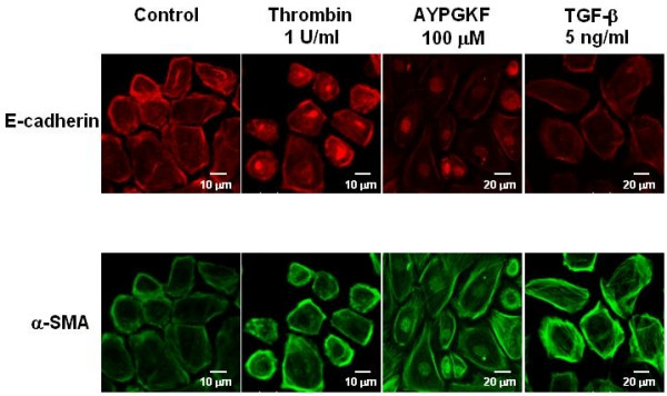
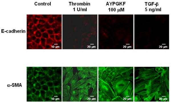
EMT was assessed by measuring the changes in each specific cell markers, E-cadherin for epithelial cell, alpha-smooth muscle actin (alpha-SMA) for myofibroblast, using primary cultured mouse alveolar epithelial cells and human lung carcinoma-derived alveolar epithelial cell line (A549 cells).
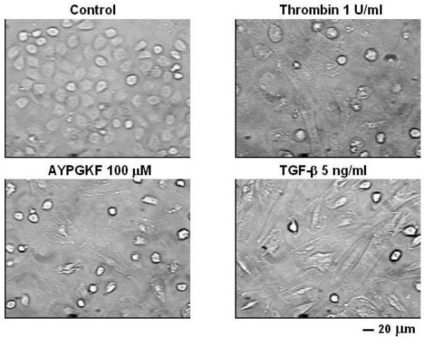
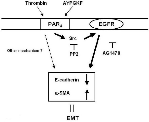
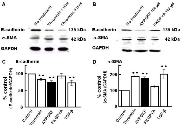
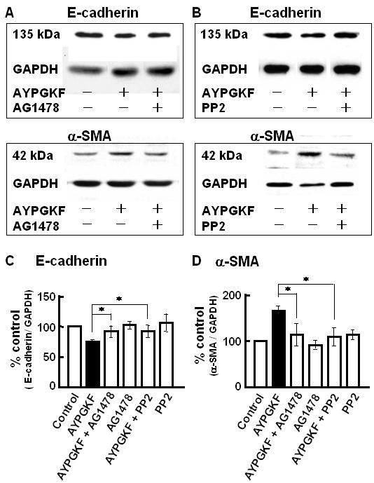
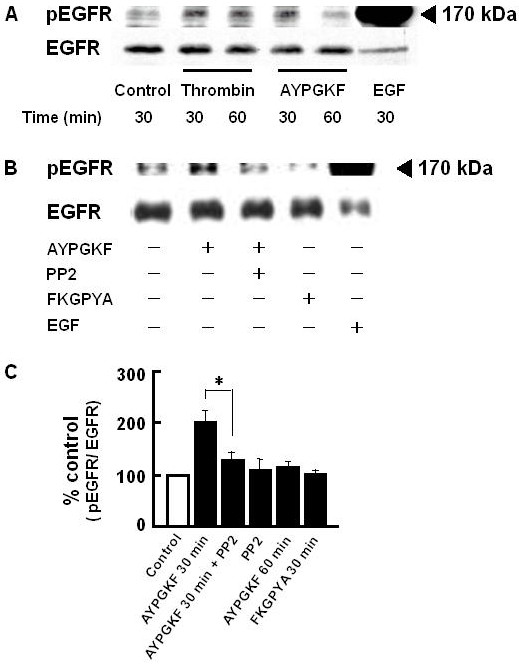
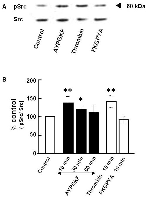
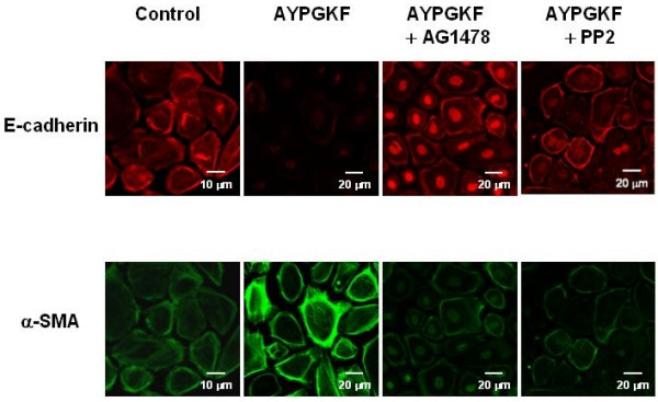
Stimulation of PAR with thrombin (1 U/ml) or a synthetic PAR4 agonist peptide (AYPGKF-NH2, 100 muM) for 72 h induced morphological changes from cobblestone-like structure to elongated shape in primary cultured alveolar epithelial cells and A549 cells. In immunocytochemical analyses of these cells, such PAR4 stimulation decreased E-cadherin-like immunoreactivity and increased alpha-SMA-like immunoreactivity, as observed with a typical EMT-inducer, tumor growth factor-beta (TGF-beta). Western blot analyses of PAR4-stimulated A549 cells also showed similar changes in expression of these EMT-related marker proteins. Such PAR4-mediated changes were attenuated by inhibitors of epidermal growth factor receptor (EGFR) kinase and Src. PAR4-mediated morphological changes in primary cultured alveolar epithelial cells were reduced in the presence of these inhibitors. PAR4 stimulation increased tyrosine phosphorylated EGFR or tyrosine phosphorylated Src level in A549 cells, and the former response being inhibited by Src inhibitor.
PAR4 stimulation of alveolar epithelial cells induced epithelial-mesenchymal transition (EMT) as monitored by cell shapes, and epithelial or myofibroblast marker at least partly through EGFR transactivation via receptor-linked Src activation.
蛋白酶激活受体(PARs;PAR1 - 4)可被凝血酶和中性粒细胞组织蛋白酶G等丝氨酸蛋白酶激活,已知其在包括纤维化在内的各种肺部疾病的发病机制中起作用。在这些PARs中,尤其是新发现的亚型PAR4,在肺中高表达。在此,我们研究了PAR4刺激是否通过促进成肌纤维细胞数量增加的肺泡上皮 - 间充质转化(EMT)在肺纤维化反应形成中发挥作用。
使用原代培养的小鼠肺泡上皮细胞和人肺癌来源的肺泡上皮细胞系(A549细胞),通过测量每种特定细胞标志物(上皮细胞的E - 钙黏蛋白和成肌纤维细胞的α - 平滑肌肌动蛋白(α - SMA))的变化来评估EMT。
用凝血酶(1 U/ml)或合成的PAR4激动剂肽(AYPGKF - NH2,100 μM)刺激PAR 72小时,可诱导原代培养的肺泡上皮细胞和A549细胞从鹅卵石样结构转变为细长形状的形态变化。在这些细胞的免疫细胞化学分析中,与典型的EMT诱导剂肿瘤生长因子 - β(TGF - β)一样,这种PAR4刺激降低了E - 钙黏蛋白样免疫反应性并增加了α - SMA样免疫反应性。对PAR4刺激的A549细胞进行的蛋白质印迹分析也显示了这些EMT相关标志物蛋白表达的类似变化。这些PAR4介导的变化被表皮生长因子受体(EGFR)激酶和Src的抑制剂减弱。在这些抑制剂存在的情况下,PAR4介导的原代培养肺泡上皮细胞的形态变化减少。PAR4刺激增加了A549细胞中酪氨酸磷酸化的EGFR或酪氨酸磷酸化的Src水平,并且前一种反应被Src抑制剂抑制。
PAR4刺激肺泡上皮细胞诱导上皮 - 间充质转化(EMT),这通过细胞形状以及上皮或成肌纤维细胞标志物来监测,至少部分是通过受体连接的Src激活介导的EGFR反式激活实现的。