Conner Ian P, Odom J Vernon, Schwartz Terry L, Mendola Janine D
Department of Neurobiology and Anatomy, West Virginia University, Morgantown, West Virginia 26505, USA.
J AAPOS. 2007 Aug;11(4):341-50. doi: 10.1016/j.jaapos.2007.01.119. Epub 2007 Apr 16.
Although previous neuroimaging efforts clearly indicate visual cortical dysfunction in adults with amblyopia, the extent of abnormalities remains unclear.
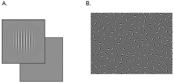
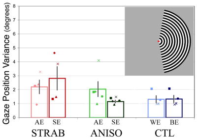
This functional magnetic resonance imaging (fMRI) study directly compared activity in visual cortex produced by monocular stimulation in 18 adults (six esotropic strabismics, six anisometropes, and six controls). Measures were made in three cortical regions-of-interest, individually defined using standard retinotopic mapping techniques in the nonamblyopic eye, corresponding to extrafoveal V1, extrafoveal V2, and the foveal representation at the occipital pole. Fixation stability was monitored and found not to differ significantly between subject groups.
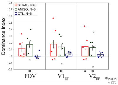
Overall results showed depressed fMRI signal magnitude for amblyopic eyes compared with sound eyes, although a few subjects did not show this trend. Assessment of the spatial extent of activation using an ocular dominance index did show significantly larger interocular differences for both strabismics and anisometropes compared with control subjects for whom eye dominance was carefully defined. In addition, both amblyopic groups showed less cortical area able to be significantly driven by either eye. The magnitude of these effects was equivalent in V1, V2, and the foveal representation, as well as between amblyopic groups. No difference was detected in the strength of signal from the nasal versus temporal retina in either amblyopic group.
Asymmetries in magnitude of monocular activation do occur in subjects with amblyopia, but these basic measures are limited in terms of sensitivity for mild to moderate amblyopia and for specificity between subtypes.
尽管先前的神经影像学研究明确表明成人弱视患者存在视觉皮层功能障碍,但异常程度仍不明确。
这项功能磁共振成像(fMRI)研究直接比较了18名成年人(6名内斜视患者、6名屈光参差患者和6名对照者)单眼刺激时视觉皮层的活动。测量在三个感兴趣的皮层区域进行,这些区域在非弱视眼中使用标准视网膜定位映射技术单独定义,分别对应于中央凹外V1、中央凹外V2以及枕极的中央凹表征。监测注视稳定性,发现各受试者组之间无显著差异。
总体结果显示,与健眼相比,弱视眼的fMRI信号强度降低,尽管有少数受试者未表现出这种趋势。使用眼优势指数评估激活的空间范围确实显示,与仔细定义了眼优势的对照受试者相比,斜视患者和屈光参差患者的两眼间差异明显更大。此外,两个弱视组中能够被任一眼显著驱动的皮层面积都较小。这些效应的强度在V1、V2和中央凹表征中以及在弱视组之间是相当的。在任何一个弱视组中,未检测到来自鼻侧视网膜与颞侧视网膜的信号强度差异。
弱视患者确实存在单眼激活强度的不对称,但这些基本测量方法在对轻度至中度弱视的敏感性以及亚型之间的特异性方面存在局限性。