Proulx Stéphanie, Bourget Jean-Michel, Gagnon Nicolas, Martel Sophie, Deschambeault Alexandre, Carrier Patrick, Giasson Claude J, Auger François A, Brunette Isabelle, Germain Lucie
Laboratoire d'Organogénèse Experiméntale, Hôpital du St-Sacrement du Centre Hospitalier Affilié Universitaire de Québec and Department of Oto-Rhino-Laryngology and Ophthalmology, Université Laval, Québec, Canada.
Mol Vis. 2007 Apr 3;13:524-33.
To optimize the growth condition of porcine corneal endothelial cells (PCEC), we evaluated the effect of coculturing with a feeder layer (irradiated 3T3 fibroblasts) with the addition of various exogenous factors, such as epidermal growth factor (EGF), nerve growth factor (NGF), bovine pituitary extract (BPE), ascorbic acid, and chondroitin sulfate, on cell proliferation, size, and morphology.
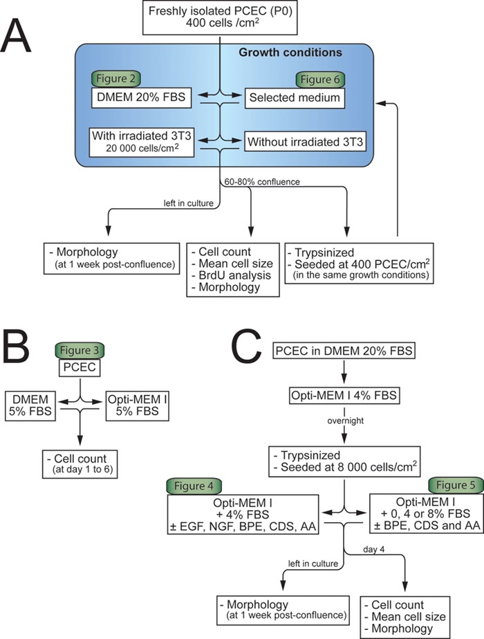
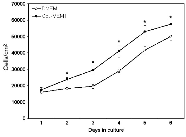
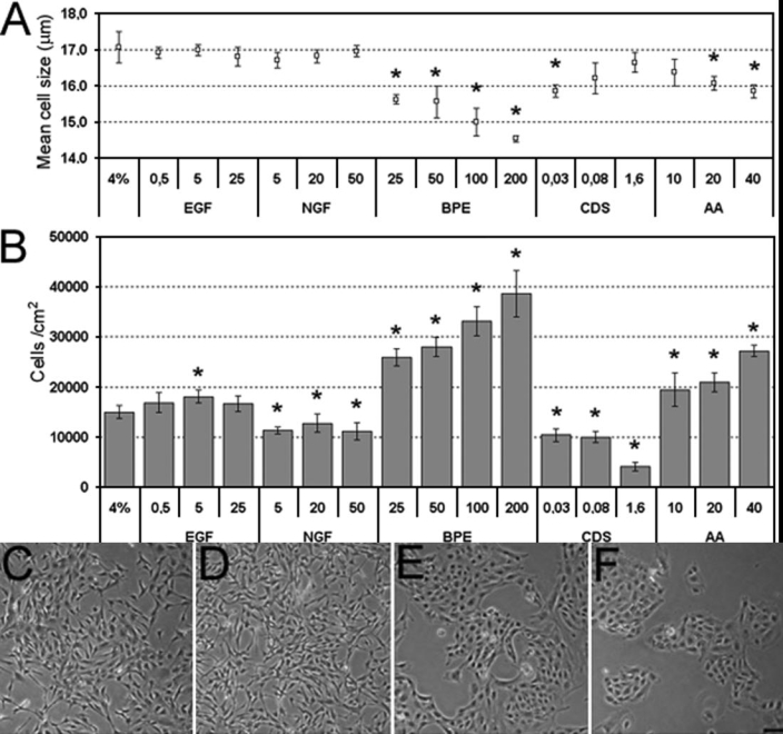
PCEC cultures were seeded at an initial cell density of 400 cells/cm(2) in the presence or absence of 20,000 murine-irradiated 3T3 fibroblast/cm(2) in the classic media Dulbecco's Modified Eagle's Medium (DMEM) supplemented with 20% fetal bovine serum (FBS). Mean cell size and bromodeoxyuridine incorporation was assessed at various passages. Growth-promoting factors were studies by seeding PCEC at 8,000 cells/cm(2) in DMEM with 20% FBS or Opti-MEM I supplemented with 4% FBS and one of the following additives: EGF (0.5, 5, 25 ng/ml), NGF (5, 20, 50 ng/ml), BPE (25, 50, 100, 200 microg/ml), ascorbic acid (10, 20, 40 microg/ml) and chondroitin sulfate (0.03, 0.08, 1.6%), alone or in combination. Cell number, size and morphology of PCEC were assessed on different cell populations. Each experiment was repeated at least twice in three sets. In some cases, cell cultures were maintained after confluence to observe post-confluence changes in cell morphology.
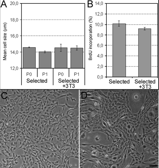
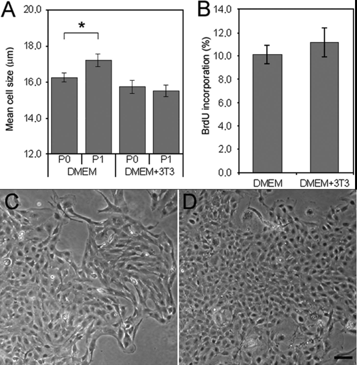
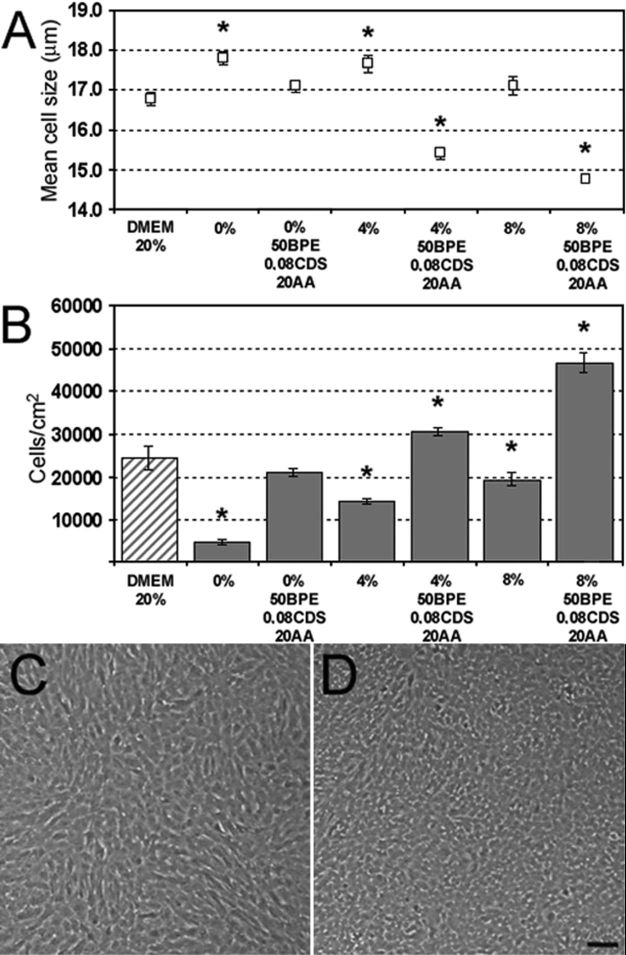
Co-cultures of PCEC grown in DMEM 20% FBS with a 3T3 feeder layer improved the preservation of small polygonal cell shape. EGF, NGF, and chondroitin sulfate did not induce proliferation above basal level nor did these additives help maintain a small size. However, chondroitin sulfate did help preserve a good morphology. BPE and ascorbic acid had dose-dependent effects on proliferation. The combination of BPE, chondroitin sulfate, and ascorbic acid significantly increased cell numbers above those achieved with serum alone. No noticeable changes were observed when PCEC were cocultured with a 3T3 feeder layer in the final selected medium.
Improvements have been made for the culture of PCEC. The final selected medium consistently allowed the growth of a contact-inhibited cell monolayer of small, polygonal-shaped cells.
为优化猪角膜内皮细胞(PCEC)的生长条件,我们评估了与饲养层(经辐照的3T3成纤维细胞)共培养,并添加各种外源性因子,如表皮生长因子(EGF)、神经生长因子(NGF)、牛垂体提取物(BPE)、抗坏血酸和硫酸软骨素,对细胞增殖、大小和形态的影响。
将PCEC培养物以400个细胞/cm²的初始细胞密度接种于含有或不含有20000个经鼠源辐照的3T3成纤维细胞/cm²的经典培养基杜氏改良 Eagle 培养基(DMEM)中,该培养基补充有20%胎牛血清(FBS)。在不同传代时评估平均细胞大小和溴脱氧尿苷掺入情况。通过将PCEC以8000个细胞/cm²接种于含有20% FBS的DMEM或补充有4% FBS的Opti-MEM I中,并添加以下添加剂之一来研究生长促进因子:EGF(0.5、5、25 ng/ml)、NGF(5、20、50 ng/ml)、BPE(25、50、100、200 μg/ml)、抗坏血酸(10、20、40 μg/ml)和硫酸软骨素(0.03、0.08、1.6%),单独或组合使用。评估不同细胞群体中PCEC的细胞数量、大小和形态。每个实验至少重复三次,每次重复至少两次。在某些情况下,汇合后维持细胞培养以观察汇合后细胞形态的变化。
在含有20% FBS的DMEM中与3T3饲养层共培养的PCEC改善了小多边形细胞形状的保持。EGF、NGF和硫酸软骨素未诱导增殖超过基础水平,这些添加剂也无助于维持小尺寸。然而,硫酸软骨素确实有助于保持良好的形态。BPE和抗坏血酸对增殖有剂量依赖性影响。BPE、硫酸软骨素和抗坏血酸的组合显著增加了细胞数量,超过了仅用血清所达到的数量。当在最终选定的培养基中PCEC与3T3饲养层共培养时,未观察到明显变化。
PCEC的培养已得到改进。最终选定的培养基始终允许生长出由小的多边形细胞组成的接触抑制细胞单层。