Frenguelli Bruno G, Wigmore Geoffrey, Llaudet Enrique, Dale Nicholas
Neurosciences Institute, Division of Pathology and Neuroscience, University of Dundee, Ninewells Hospital, Dundee, UK.
J Neurochem. 2007 Jun;101(5):1400-13. doi: 10.1111/j.1471-4159.2006.04425.x.
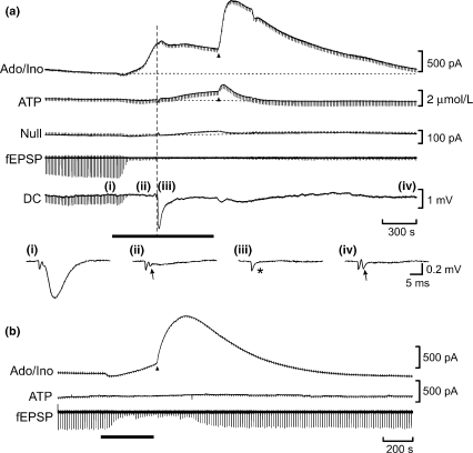
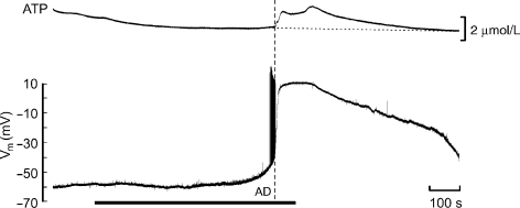
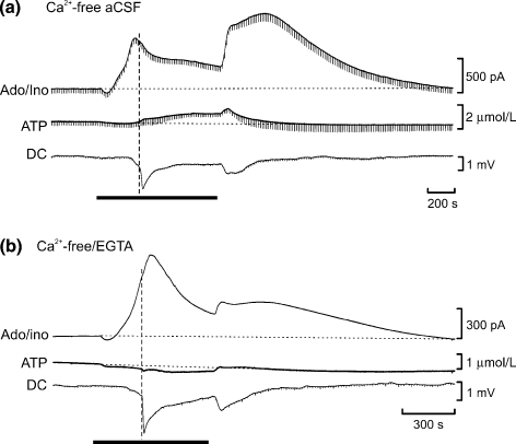
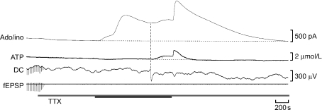
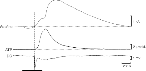
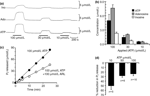
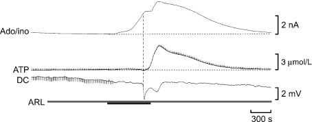
Adenosine is well known to be released during cerebral metabolic stress and is believed to be neuroprotective. ATP release under similar circumstances has been much less studied. We have now used biosensors to measure and compare in real time the release of ATP and adenosine during in vitro ischaemia in hippocampal slices. ATP release only occurred following the anoxic depolarisation, whereas adenosine release was apparent almost immediately after the onset of ischaemia. ATP release required extracellular Ca2+. By contrast adenosine release was enhanced by removal of extracellular Ca2+, whilst TTX had no effect on either ATP release or adenosine release. Blockade of ionotropic glutamate receptors substantially enhanced ATP release, but had only a modest effect on adenosine release. Carbenoxolone, an inhibitor of gap junction hemichannels, also greatly enhanced ischaemic ATP release, but had little effect on adenosine release. The ecto-ATPase inhibitor ARL 67156, whilst modestly enhancing the ATP signal detected during ischaemia, had no effect on adenosine release. Adenosine release during ischaemia was reduced by pretreatment with homosysteine thiolactone suggesting an intracellular origin. Adenosine transport inhibitors did not inhibit adenosine release, but instead they caused a twofold increase of release. Our data suggest that ATP and adenosine release during ischaemia are for the most part independent processes with distinct underlying mechanisms. These two purines will consequently confer temporally distinct influences on neuronal and glial function in the ischaemic brain.
众所周知,腺苷在脑代谢应激期间会释放,并且被认为具有神经保护作用。在类似情况下ATP的释放则研究得少得多。我们现在使用生物传感器实时测量和比较海马切片体外缺血期间ATP和腺苷的释放。ATP仅在缺氧去极化后释放,而腺苷释放几乎在缺血开始后立即明显出现。ATP的释放需要细胞外Ca2+。相比之下,去除细胞外Ca2+可增强腺苷释放,而TTX对ATP释放或腺苷释放均无影响。阻断离子型谷氨酸受体可显著增强ATP释放,但对腺苷释放仅有适度影响。缝隙连接半通道抑制剂羧苄青霉素也可大大增强缺血性ATP释放,但对腺苷释放影响不大。胞外ATP酶抑制剂ARL 67156虽然适度增强了缺血期间检测到的ATP信号,但对腺苷释放无影响。同型半胱氨酸硫内酯预处理可减少缺血期间的腺苷释放,提示其起源于细胞内。腺苷转运抑制剂并不抑制腺苷释放,反而使其释放增加两倍。我们的数据表明,缺血期间ATP和腺苷的释放大多是独立过程,其潜在机制不同。因此,这两种嘌呤将对缺血性脑内的神经元和胶质细胞功能产生不同时间的影响。