Lee Yoon Jung, Kang Sung Min, Kang Il Bong
Department of Ophthalmology, Hanyang University, School of Medicine, Hanyang University Guri Hospital, Gyeonggi-do, Korea.
Korean J Ophthalmol. 2007 Mar;21(1):61-4. doi: 10.3341/kjo.2007.21.1.61.
To report a case of acute angle-closure glaucoma resulting from spontaneous hemorrhagic retinal detachment.
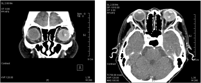
An 81-year-old woman visited our emergency room for severe ocular pain and vision loss in her left eye. Her intraocular pressures (IOPs) were 14 mmHg in the right eye and 58 mmHg in the left eye. Her visual acuity was 0.4 in the right eye but she had no light perception in the left eye. The left anterior chamber depth was shallow and gonioscopy of the left eye showed a closed angle. In comparison, the right anterior chamber depth was normal and showed a wide, open angle. Computed tomography and ultrasonography demonstrated retinal detachment due to subretinal hemorrhage. After systemic and topical antiglaucoma medications failed to relieve her intractable severe ocular pain, she underwent enucleation.
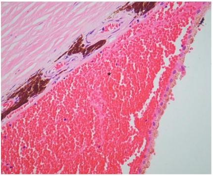
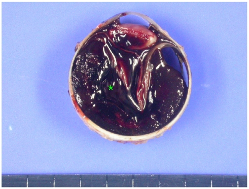
The ocular pathology specimen showed that a large subretinal hemorrhage caused retinal detachment and pushed displaced the lens-iris diaphragm, resulting in secondary angle-closure glaucoma.
Prolonged anticoagulant therapy may cause hemorrhagic retinal detachment and secondary angle-closure glaucoma. If medical therapy fails to relieve pain or if there is suspicion of an intraocular tumor, enucleation should be considered as a therapeutic option.
报告一例因自发性出血性视网膜脱离导致的急性闭角型青光眼病例。
一名81岁女性因左眼严重眼痛和视力丧失前来我院急诊室就诊。她的右眼眼压(IOP)为14 mmHg,左眼眼压为58 mmHg。她的右眼视力为0.4,但左眼无光感。左眼前房深度浅,左眼房角镜检查显示房角关闭。相比之下,右眼前房深度正常,房角宽且开放。计算机断层扫描和超声检查显示视网膜下出血导致视网膜脱离。在全身和局部抗青光眼药物治疗未能缓解其顽固性严重眼痛后,她接受了眼球摘除术。
眼部病理标本显示,大量视网膜下出血导致视网膜脱离,并推动晶状体虹膜隔移位,导致继发性闭角型青光眼。
长期抗凝治疗可能导致出血性视网膜脱离和继发性闭角型青光眼。如果药物治疗无法缓解疼痛或怀疑有眼内肿瘤,应考虑将眼球摘除术作为一种治疗选择。