Massey James M, Amps Jeremy, Viapiano Mariano S, Matthews Russell T, Wagoner Michelle R, Whitaker Christopher M, Alilain Warren, Yonkof Alicia L, Khalyfa Abdelnaby, Cooper Nigel G F, Silver Jerry, Onifer Stephen M
School of Medicine, University of Louisville, Louisville, KY 40292, USA.
Exp Neurol. 2008 Feb;209(2):426-45. doi: 10.1016/j.expneurol.2007.03.029. Epub 2007 Apr 12.
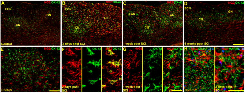
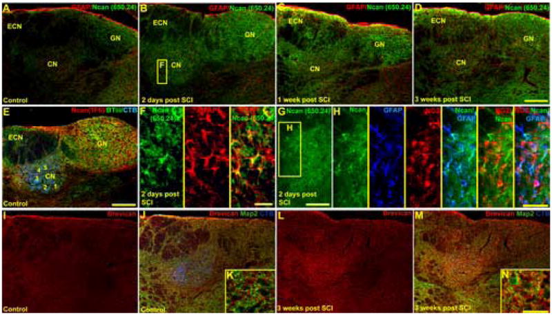
Increased chondroitin sulfate proteoglycan (CSPG) expression in the vicinity of a spinal cord injury (SCI) is a primary participant in axonal regeneration failure. However, the presence of similar increases of CSPG expression in denervated synaptic targets well away from the primary lesion and the subsequent impact on regenerating axons attempting to approach deafferented neurons have not been studied. Constitutively expressed CSPGs within the extracellular matrix and perineuronal nets of the adult rat dorsal column nuclei (DCN) were characterized using real-time PCR, Western blot analysis and immunohistochemistry. We show for the first time that by 2 days and through 3 weeks following SCI, the levels of NG2, neurocan and brevican associated with reactive glia throughout the DCN were dramatically increased throughout the DCN despite being well beyond areas of trauma-induced blood brain barrier breakdown. Importantly, regenerating axons from adult sensory neurons microtransplanted 2 weeks following SCI between the injury site and the DCN were able to regenerate rapidly within white matter (as shown previously by Davies et al. [Davies, S.J., Goucher, D.R., Doller, C., Silver, J., 1999. Robust regeneration of adult sensory axons in degenerating white matter of the adult rat spinal cord. J. Neurosci. 19, 5810-5822]) but were unable to enter the denervated DCN. Application of chondroitinase ABC or neurotrophin-3-expressing lentivirus in the DCN partially overcame this inhibition. When the treatments were combined, entrance by regenerating axons into the DCN was significantly augmented. These results demonstrate both an additional challenge and potential treatment strategy for successful functional pathway reconstruction after SCI.
脊髓损伤(SCI)附近硫酸软骨素蛋白聚糖(CSPG)表达增加是轴突再生失败的主要原因。然而,在远离原发性损伤的失神经支配突触靶点中CSPG表达是否有类似增加以及对试图接近去传入神经元的再生轴突的后续影响尚未得到研究。使用实时PCR、蛋白质印迹分析和免疫组织化学对成年大鼠背柱核(DCN)细胞外基质和神经元周围网中组成性表达的CSPG进行了表征。我们首次表明,在SCI后2天至3周内,尽管DCN远超出创伤诱导的血脑屏障破坏区域,但整个DCN中与反应性胶质细胞相关的NG2、神经聚糖和短蛋白聚糖水平显著增加。重要的是,SCI后2周在损伤部位和DCN之间微移植的成年感觉神经元的再生轴突能够在白质中快速再生(如Davies等人先前所示[Davies, S.J., Goucher, D.R., Doller, C., Silver, J., 1999. Robust regeneration of adult sensory axons in degenerating white matter of the adult rat spinal cord. J. Neurosci. 19, 5810-5822]),但无法进入失神经支配的DCN。在DCN中应用软骨素酶ABC或表达神经营养因子-3的慢病毒部分克服了这种抑制作用。当联合使用这些治疗方法时,再生轴突进入DCN的情况显著增加。这些结果证明了SCI后成功进行功能通路重建面临的额外挑战和潜在治疗策略。