Brand Christine, Schaeffel Frank, Feldkaemper Marita Pauline
Section for Neurobiology of the Eye, University Eye Hospital Tuebingen, Tuebingen, Germany.
Mol Vis. 2007 Jun 18;13:920-32.
The development of myopia is controlled by still largely unknown retinal signals. The aim of this study was to investigate the changes in retinal mRNA expression after different periods of visual deprivation in mice, while controlling for retinal illuminance.
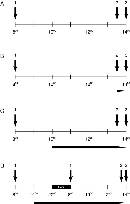
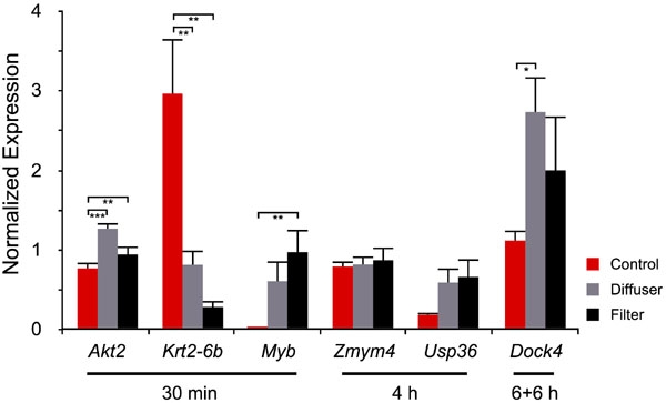
Each group consisted of three male C57BL/6 mice. Treatment periods were 30 min, 4 h, and 6+6 h. High spatial frequencies were filtered from the retinal image by frosted diffusers over one eye while the fellow eyes were covered by clear neutral density (ND) filters that exhibited similar light attenuating properties (0.1 log units) as the diffusers. For the final 30 min of the respective treatment period mice were individually placed in a clear Perspex cylinder that was positioned in the center of a rotating (60 degrees) large drum. The inside of the drum was covered with a 0.1 cyc/degree vertical square wave grating. This visual environment was chosen to standardize illuminances and contrasts seen by the mice. Labeled cRNA was prepared and hybridized to Affymetrix GeneChip Mouse Genome 430 2.0 arrays. Alterations in mRNA expression levels of candidate genes with potential biological relevance were confirmed by semi-quantitative real-time reverse transcription polymerase chain reaction (RT-PCR).
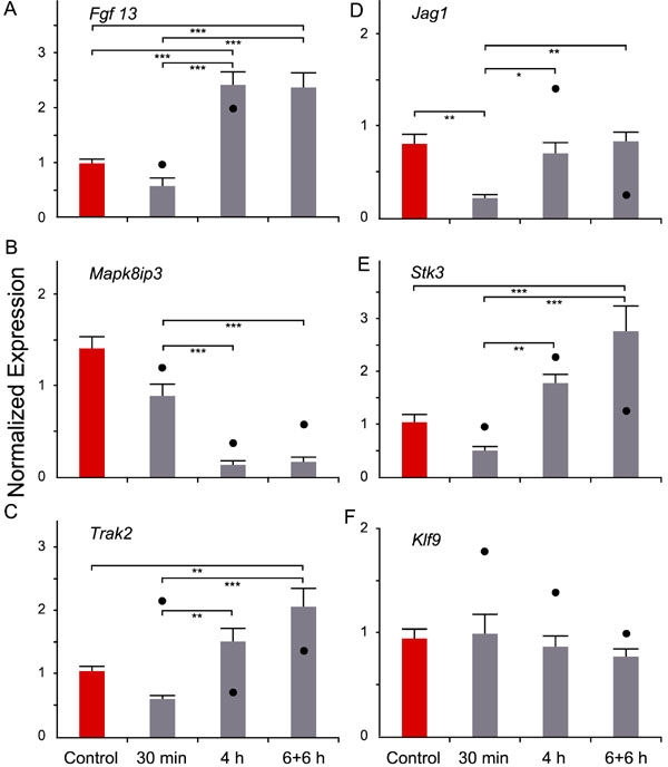
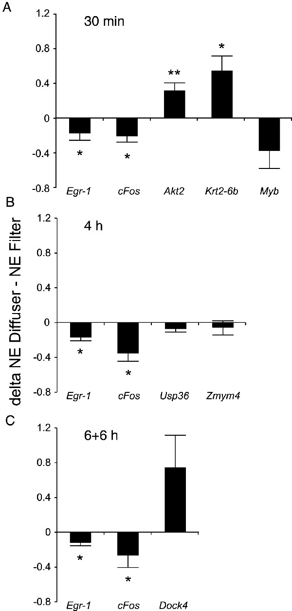
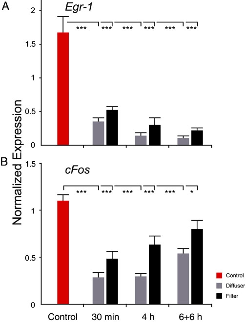
In all groups, Egr-1 mRNA expression was reduced in diffuser-treated eyes. Furthermore, the degradation of the spatial frequency spectrum also changed the cFos mRNA level, with reduced expression after 4 h of diffuser treatment. Other interesting candidates were Akt2, which was up-regulated after 30 min of deprivation and Mapk8ip3, a neuron specific JNK binding and scaffolding protein that was temporally regulated in the diffuser-treated eyes only.
The microarray analysis demonstrated a pattern of differential transcriptional changes, even though differences in the retinal images were restricted to spatial features. The candidate genes may provide further insight into the biochemical short-term changes following retinal image degradation in mice. Because deprivation of spatial vision leads to increased eye growth and myopia in both animals and humans, it is believed some of the identified genes play a role in myopia development.
近视的发展仍在很大程度上受未知的视网膜信号控制。本研究的目的是在控制视网膜照度的同时,研究小鼠在不同时间段视觉剥夺后视网膜mRNA表达的变化。
每组由三只雄性C57BL/6小鼠组成。治疗期分别为30分钟、4小时和6 + 6小时。用磨砂扩散器对一只眼睛的视网膜图像进行高空间频率滤波,而另一只眼睛则覆盖有与扩散器具有相似光衰减特性(0.1对数单位)的透明中性密度(ND)滤光片。在各自治疗期的最后30分钟,将小鼠单独放置在一个透明的有机玻璃圆筒中,该圆筒位于一个旋转(60度)的大鼓的中心。鼓的内部覆盖有一个0.1周/度的垂直方波光栅。选择这种视觉环境来标准化小鼠所看到的照度和对比度。制备标记的cRNA并与Affymetrix GeneChip Mouse Genome 430 2.0阵列杂交。通过半定量实时逆转录聚合酶链反应(RT-PCR)确认具有潜在生物学相关性的候选基因mRNA表达水平的变化。
在所有组中,扩散器处理的眼睛中Egr-1 mRNA表达降低。此外,空间频谱的退化也改变了cFos mRNA水平,扩散器处理4小时后表达降低。其他有趣的候选基因是Akt2,其在剥夺30分钟后上调,以及Mapk8ip3,一种仅在扩散器处理的眼睛中受到时间调节的神经元特异性JNK结合和支架蛋白。
微阵列分析显示了差异转录变化的模式,尽管视网膜图像的差异仅限于空间特征。候选基因可能为进一步了解小鼠视网膜图像退化后的生化短期变化提供线索。由于空间视觉剥夺会导致动物和人类的眼轴增长和近视增加,可以认为一些已鉴定的基因在近视发展中起作用。