Riddell Nina, Faou Pierre, Murphy Melanie, Giummarra Loretta, Downs Rachael A, Rajapaksha Harinda, Crewther Sheila G
Department of Psychology and Counselling, School of Psychology and Public Health, La Trobe University, Melbourne, VIC, Australia.
Department of Biochemistry and Genetics, La Trobe Institute for Molecular Sciences, La Trobe University, Melbourne, VIC, Australia.
Mol Vis. 2017 Dec 5;23:872-888. eCollection 2017.
Microarray and RNA sequencing studies in the chick model of early optically induced refractive error have implicated thousands of genes, many of which have also been linked to ocular pathologies in humans, including age-related macular degeneration (AMD), choroidal neovascularization, glaucoma, and cataract. These findings highlight the potential relevance of the chick model to understanding both refractive error development and the progression to secondary pathological complications. The present study aimed to determine whether proteomic responses to early optical defocus in the chick share similarities with these transcriptome-level changes, particularly in terms of dysregulation of pathology-related molecular processes.
Chicks were assigned to a lens condition (monocular +10 D [diopters] to induce hyperopia, -10 D to induce myopia, or no lens) on post-hatch day 5. Biometric measures were collected following a further 6 h and 48 h of rearing. The retina/RPE was then removed and prepared for liquid chromatography-electrospray ionization-tandem mass spectrometry (LC-ESI-MS/MS) on an LTQ-Orbitrap Elite. Raw data were processed using MaxQuant, and differentially abundant proteins were identified using moderated tests (fold change ≥1.5, Benjamini-Hochberg adjusted p<0.05). These differentially abundant proteins were compared with the genes and proteins implicated in previous exploratory transcriptome and proteomic studies of refractive error, as well as the genes and proteins linked to the ocular pathologies listed above for which myopia or hyperopia are risk factors. Finally, gene set enrichment analysis (GSEA) was used to assess whether gene sets from the Human Phenotype Ontology database were enriched in the lens groups relative to the no lens groups, and at the top or bottom of the protein data ranked by Spearman's correlation with refraction at 6 and 48 h.
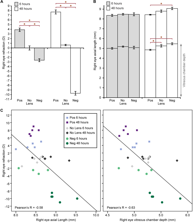
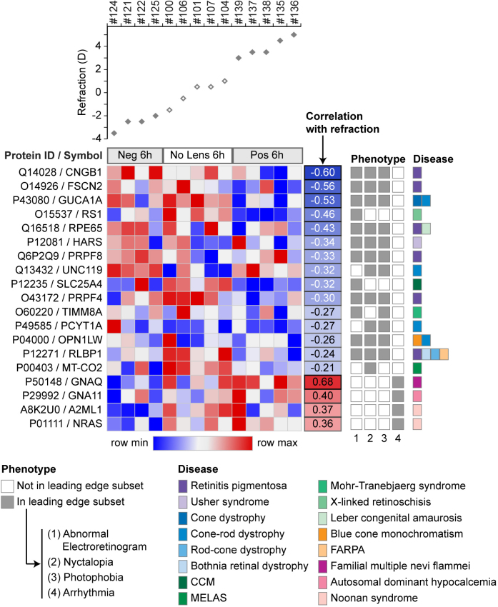
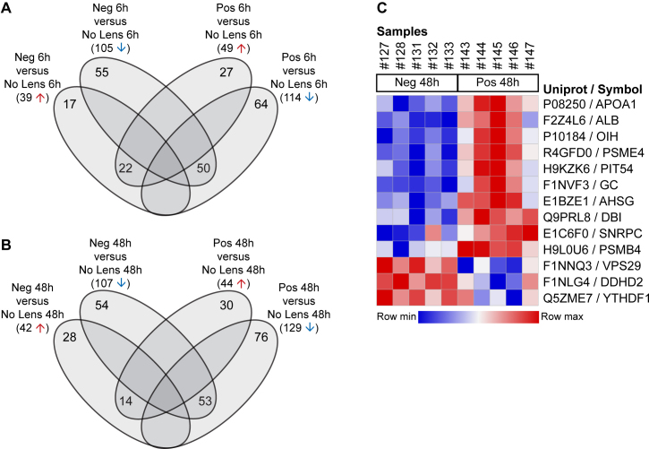
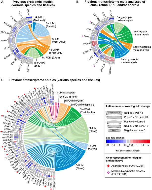
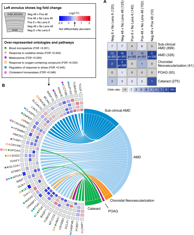
Refractive errors of -2.63 D ± 0.31 D (mean ± standard error, SE) and 3.90 D ± 0.37 D were evident in the negative and positive lens groups, respectively, at 6 h. By 48 h, refractive compensation to both lens types was almost complete (negative lens -9.70 D ± 0.41 D, positive lens 7.70 D ± 0.44 D). More than 140 differentially abundant proteins were identified in each lens group relative to the no lens controls at both time points. No proteins were differentially abundant between the negative and positive lens groups at 6 h, and 13 were differentially abundant at 48 h. As there was substantial overlap in the proteins implicated across the six comparisons, a total of 390 differentially abundant proteins were identified. Sixty-five of these 390 proteins had previously been implicated in transcriptome studies of refractive error animal models, and 42 had previously been associated with AMD, choroidal neovascularization, glaucoma, and/or cataract in humans. The overlap of differentially abundant proteins with AMD-associated genes and proteins was statistically significant for all conditions (Benjamini-Hochberg adjusted p<0.05), with over-representation analysis implicating ontologies related to oxidative stress, cholesterol homeostasis, and melanin biosynthesis. GSEA identified significant enrichment of genes associated with abnormal electroretinogram, photophobia, and nyctalopia phenotypes in the proteins negatively correlated with ocular refraction across the lens groups at 6 h. The implicated proteins were primarily linked to photoreceptor dystrophies and mitochondrial disorders in humans.
Optical defocus in the chicks induces rapid changes in the abundance of many proteins in the retina/RPE that have previously been linked to inherited and age-related ocular pathologies in humans. Similar changes have been identified in a meta-analysis of chick refractive error transcriptome studies, highlighting the chick as a model for the study of optically induced stress with possible relevance to understanding the development of a range of pathological states in humans.
在早期光学诱导屈光不正的雏鸡模型中进行的微阵列和RNA测序研究涉及数千个基因,其中许多基因也与人类眼部疾病有关,包括年龄相关性黄斑变性(AMD)、脉络膜新生血管形成、青光眼和白内障。这些发现凸显了雏鸡模型在理解屈光不正发展以及继发病理并发症进展方面的潜在相关性。本研究旨在确定雏鸡对早期光学离焦的蛋白质组反应是否与这些转录组水平的变化具有相似性,特别是在与病理相关的分子过程失调方面。
在雏鸡孵化后第5天,将其分为晶状体条件组(单眼+10屈光度[D]诱导远视,-10 D诱导近视,或无晶状体)。在进一步饲养6小时和48小时后收集生物测量数据。然后取出视网膜/视网膜色素上皮(RPE),在LTQ-Orbitrap Elite上进行液相色谱-电喷雾电离-串联质谱(LC-ESI-MS/MS)分析。原始数据使用MaxQuant进行处理,使用适度检验鉴定差异丰富的蛋白质(倍数变化≥1.5,Benjamini-Hochberg校正p<0.05)。将这些差异丰富的蛋白质与先前屈光不正探索性转录组和蛋白质组研究中涉及的基因和蛋白质进行比较,以及与上述近视或远视为危险因素的眼部疾病相关的基因和蛋白质进行比较。最后,使用基因集富集分析(GSEA)评估来自人类表型本体数据库的基因集在晶状体组中相对于无晶状体组是否富集,以及在与6小时和48小时屈光力的Spearman相关性排序的蛋白质数据的顶部或底部。
在6小时时,负性和正性晶状体组的屈光不正分别为-2.63 D±0.31 D(平均值±标准误,SE)和3.90 D±0.37 D。到48小时时,两种晶状体类型的屈光补偿几乎完成(负性晶状体-9.70 D±0.41 D,正性晶状体7.70 D±0.44 D)。在两个时间点,相对于无晶状体对照组,每个晶状体组中鉴定出超过140种差异丰富的蛋白质。在6小时时,负性和正性晶状体组之间没有差异丰富的蛋白质,在48小时时有13种差异丰富的蛋白质。由于在六次比较中涉及的蛋白质有大量重叠,共鉴定出390种差异丰富的蛋白质。这390种蛋白质中有65种先前已涉及屈光不正动物模型的转录组研究,42种先前与人的AMD、脉络膜新生血管形成、青光眼和/或白内障相关。差异丰富的蛋白质与AMD相关基因和蛋白质的重叠在所有条件下均具有统计学意义(Benjamini-Hochberg校正p<0.05),过度表达分析涉及与氧化应激、胆固醇稳态和黑色素生物合成相关的本体。GSEA在6小时时鉴定出与眼屈光力呈负相关的蛋白质中与异常视网膜电图、畏光和夜盲表型相关的基因显著富集。所涉及的蛋白质主要与人类的光感受器营养不良和线粒体疾病有关。
雏鸡中的光学离焦诱导视网膜/RPE中许多蛋白质丰度的快速变化,这些蛋白质先前已与人类的遗传性和年龄相关性眼部疾病相关。在雏鸡屈光不正转录组研究的荟萃分析中也发现了类似的变化,凸显了雏鸡作为光学诱导应激研究模型的作用,可能与理解人类一系列病理状态的发展相关。