Martínez-Navarrete Gema C, Martín-Nieto José, Esteve-Rudd Julián, Angulo Antonia, Cuenca Nicolás
Departamento de Fisiología, Genética y Microbiología, Facultad de Ciencias, Universidad de Alicante, Alicante, Spain.
Mol Vis. 2007 Jun 18;13:949-61.
Alpha-synuclein is a Parkinson's disease-linked protein of ubiquitous expression in the central nervous system. It has a proposed role in the modulation of neurotransmission and synaptic function. This study was aimed at analyzing expression of the alpha-synuclein gene in the normal retina, and characterizing its pattern of distribution in the different retinal cell types and layers in a variety of vertebrates, ranging from fish to humans.
Reverse transcriptase-polymerase chain reaction and immunoblotting were used to assess alpha-synuclein expression at both mRNA and protein levels. Its retinal distribution profile was characterized by immunohistochemical methods. With this purpose, retinal sections were analyzed under fluorescent confocal microscopy using specific antibodies against alpha-synuclein, alone and in double or triple combinations with a set of antibodies to molecular markers for the distinct retinal neuronal types. Also, synaptophysin was used as a marker for synaptic vesicles in the retina.
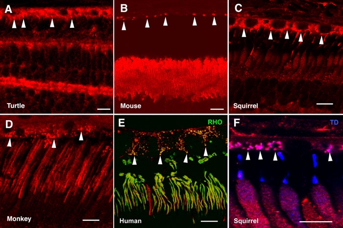
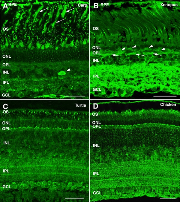
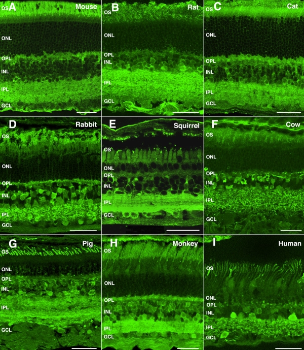
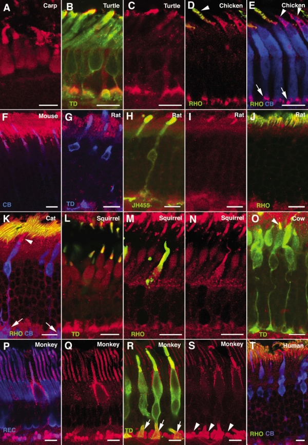
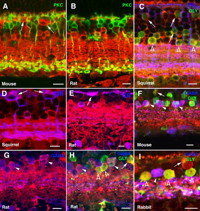
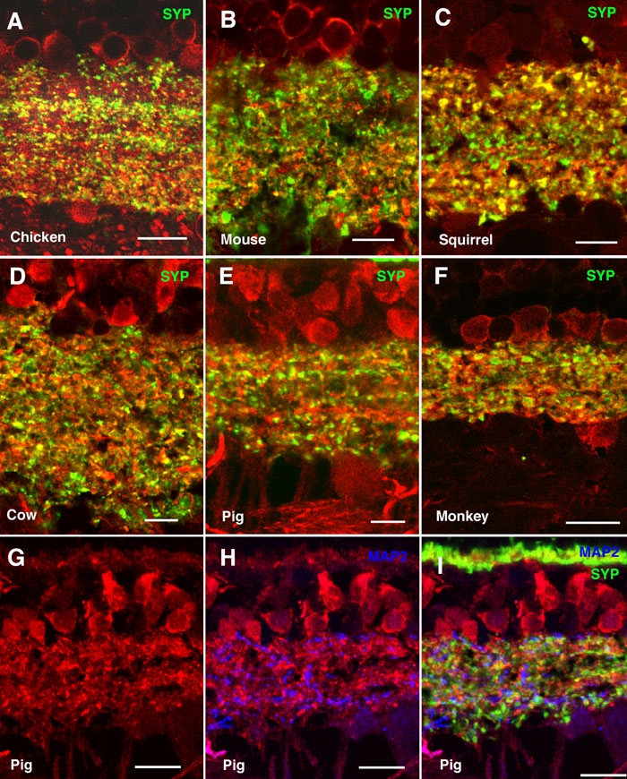
Alpha-synuclein mRNA and protein were expressed by both retinal pigment epithelium (RPE) and neural retinal cells. The pattern of alpha-synuclein distribution in the retina was quite consistent across all vertebrate species examined. A strong immunoreactivity was found in the outer segments (OS) of photoreceptors and in their axon terminals (cone pedicles and rod spherules) in the outer plexiform layer (OPL) of the retina. Alpha-synuclein was also present in rod and cone bipolar cells, as well as in GABAergic and glycinergic amacrines, distributing along a complex plexus throughout the inner plexiform layer (IPL). Additionally, colocalization was found between alpha-synuclein and synaptophysin at presynaptic terminals of the retina. Alpha-synuclein-positive phagosome-like structures were observed in the cytoplasm of RPE cells.
An involvement of alpha-synuclein can be postulated in neurotransmission at axon terminals of photoreceptors in the OPL, and at presynaptic endings of bipolar and amacrine cells in the IPL. As well, this protein could have a role in the function as well as the maintenance of photoreceptor OS. Alpha-synuclein contained in RPE cells should derive not only from protein expression by this cell type, but also from their phagocytosis of OS disc membranes.
α-突触核蛋白是一种与帕金森病相关的蛋白质,在中枢神经系统中广泛表达。它在神经传递和突触功能的调节中具有一定作用。本研究旨在分析α-突触核蛋白基因在正常视网膜中的表达情况,并确定其在从鱼类到人类等多种脊椎动物不同视网膜细胞类型和层次中的分布模式。
采用逆转录聚合酶链反应和免疫印迹法评估α-突触核蛋白在mRNA和蛋白质水平的表达。通过免疫组织化学方法确定其在视网膜中的分布特征。为此,使用针对α-突触核蛋白的特异性抗体,以及与一组针对不同视网膜神经元类型分子标记物的抗体进行单染、双染或三染,在荧光共聚焦显微镜下分析视网膜切片。此外,使用突触素作为视网膜中突触小泡的标记物。
视网膜色素上皮(RPE)细胞和神经视网膜细胞均表达α-突触核蛋白mRNA和蛋白质。在所检查的所有脊椎动物物种中,α-突触核蛋白在视网膜中的分布模式相当一致。在视网膜外丛状层(OPL)的光感受器外段(OS)及其轴突终末(视锥小足和视杆小球)中发现强烈的免疫反应性。α-突触核蛋白也存在于视杆和视锥双极细胞以及γ-氨基丁酸能和甘氨酸能无长突细胞中,沿着复杂的神经丛分布于整个内丛状层(IPL)。此外,在视网膜的突触前终末发现α-突触核蛋白与突触素共定位。在RPE细胞的细胞质中观察到α-突触核蛋白阳性的吞噬体样结构。
可以推测α-突触核蛋白参与了OPL中光感受器轴突终末以及IPL中双极细胞和无长突细胞突触前末梢的神经传递。此外,这种蛋白质可能在光感受器OS的功能以及维持中发挥作用。RPE细胞中含有的α-突触核蛋白不仅应源于该细胞类型的蛋白质表达,还应源于其对OS盘膜的吞噬作用。