Yang Li-ping, Zhu Xiu-an, Tso Mark O M
Peking University Eye Center, Peking University Third Hospital, Peking University, Beijing, PR China.
Mol Vis. 2007 Jul 9;13:1083-93.
To elucidate the inhibitory effect of minocycline and sulforaphane on lipopolysaccharide (LPS)-induced retinal microglial activation and the mechanisms through which they exerted their inhibitory effects.
Primary retinal microglial cultures were exposed to LPS with or without minocycline and sulforaphane. The mRNA expression of monocyte chemotactic protein (MCP)-1, MCP-3, macrophage inflammatory protein (MIP)-1alpha, MIP-1beta, eotaxin, regulated upon activation normal T-cell expressed and secreted (RANTES) protein, and interleukin (IL)-10 were examined by reverse transcription polymerase chain reaction (RT-PCR) assay. The mRNA expression of inducible nitric oxide synthase (iNOS) and subsequent nitric oxide (NO) production were examined by RT-PCR assay and Griess reagent assay. Protein expression of the p65 subunit of nuclear factor-kappaB (NF-kappaB) and p-p38, p-p44/42 and p-JNK mitogen-activated protein kinases (MAPKs) were examined by Western blot and immunofluorescent analysis.
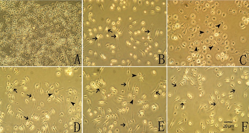
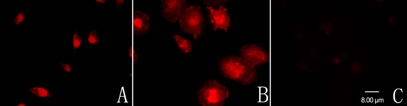
Cultured retinal microglial cells were activated following exposure to LPS. The mRNA expression and protein production of eotaxin, RANTES, and IL-10 and the mRNA expression of iNOS and subsequent NO production were upregulated. The protein expression of p-p38, p-JNK, and the p65 subunit of NF-kappaB were also upregulated. However, the protein expression of p-p44/42 was not significantly changed. Pretreatment with minocycline or sulforaphane for 1 h before LPS administration inhibited LPS-induced microglial morphological change and inhibited LPS-induced upregulation of p-p38, but had no effect on the expression of p-p44/42, p-JNK, and the p65 subunit of NF-kappaB.
Minocycline and sulforaphane inhibited LPS-induced retinal microglial activation, Western blot and immunofluorescent studies showed decreased p-p38 MAPK expression. We suggested that the inhibitory effect of minocycline and sulforaphane was partly through a p38 MAPK-dependent mechanism.
阐明米诺环素和萝卜硫素对脂多糖(LPS)诱导的视网膜小胶质细胞活化的抑制作用及其发挥抑制作用的机制。
原代视网膜小胶质细胞培养物在有或无米诺环素和萝卜硫素的情况下暴露于LPS。通过逆转录聚合酶链反应(RT-PCR)检测单核细胞趋化蛋白(MCP)-1、MCP-3、巨噬细胞炎性蛋白(MIP)-1α、MIP-1β、嗜酸性粒细胞趋化因子、活化正常T细胞表达和分泌的调节蛋白(RANTES)以及白细胞介素(IL)-10的mRNA表达。通过RT-PCR检测和Griess试剂检测诱导型一氧化氮合酶(iNOS)的mRNA表达及随后的一氧化氮(NO)生成。通过蛋白质印迹和免疫荧光分析检测核因子κB(NF-κB)p65亚基以及磷酸化p38、磷酸化p44/42和磷酸化JNK丝裂原活化蛋白激酶(MAPK)的蛋白表达。
培养的视网膜小胶质细胞在暴露于LPS后被激活。嗜酸性粒细胞趋化因子、RANTES和IL-10的mRNA表达及蛋白生成以及iNOS的mRNA表达及随后的NO生成均上调。磷酸化p38、磷酸化JNK以及NF-κB p