• 文献检索
  • 文档翻译
  • 深度研究
  • 学术资讯
  • Suppr Zotero 插件Zotero 插件
  • 邀请有礼
  • 套餐&价格
  • 历史记录
应用&插件
Suppr Zotero 插件Zotero 插件浏览器插件Mac 客户端Windows 客户端微信小程序
定价
高级版会员购买积分包购买API积分包
服务
文献检索文档翻译深度研究API 文档MCP 服务
关于我们
关于 Suppr公司介绍联系我们用户协议隐私条款
关注我们

Suppr 超能文献

核心技术专利:CN118964589B侵权必究
粤ICP备2023148730 号-1Suppr @ 2026

文献检索

告别复杂PubMed语法,用中文像聊天一样搜索,搜遍4000万医学文献。AI智能推荐,让科研检索更轻松。

立即免费搜索

文件翻译

保留排版,准确专业,支持PDF/Word/PPT等文件格式,支持 12+语言互译。

免费翻译文档

深度研究

AI帮你快速写综述,25分钟生成高质量综述,智能提取关键信息,辅助科研写作。

立即免费体验

一种分析乳腺癌患者电阻抗光谱数据的方法。

A method for analyzing electrical impedance spectroscopy data from breast cancer patients.

作者信息

Kim Bong Seok, Isaacson David, Xia Hongjun, Kao Tzu-Jen, Newell Jonathan C, Saulnier Gary J

机构信息

Department of Biomedical Engineering, Rensselaer Polytechnic Institute, Troy, NY 12180, USA.

出版信息

Physiol Meas. 2007 Jul;28(7):S237-46. doi: 10.1088/0967-3334/28/7/S17. Epub 2007 Jun 26.

DOI:10.1088/0967-3334/28/7/S17
PMID:17664638
原文链接:https://pmc.ncbi.nlm.nih.gov/articles/PMC2435084/
Abstract

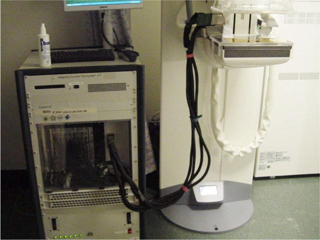
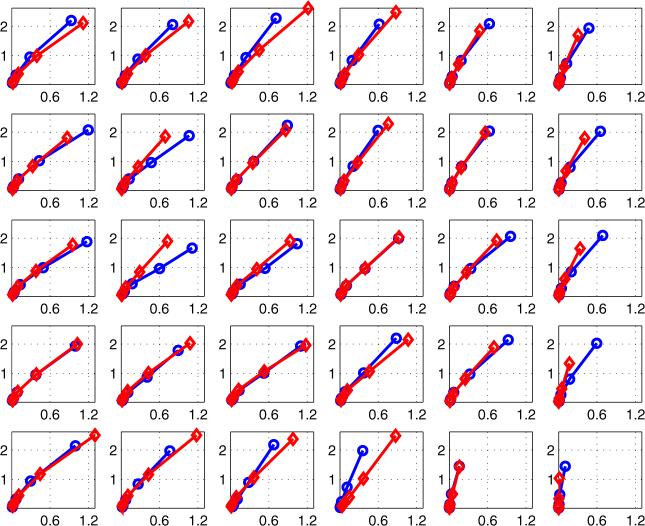
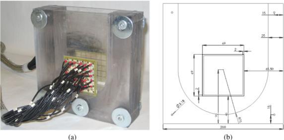
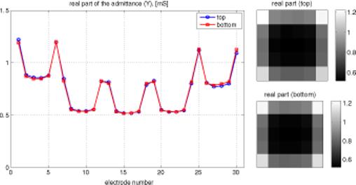
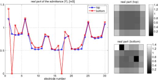
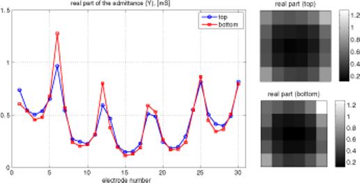
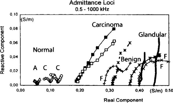
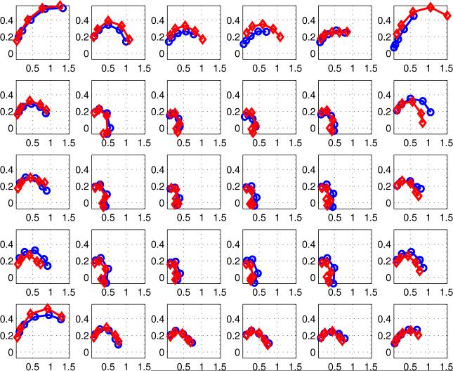
Research on freshly-excised malignant breast tissues and surrounding normal tissues in an in vitro impedance cell has shown that breast tumors have different conductivity and permittivity from normal or non-malignant tissues. This contrast may provide a basis for breast cancer detection using electrical impedance imaging. This paper describes a procedure for collecting electrical impedance spectroscopy data simultaneously and in register with tomosynthesis data from patients. We describe the methods used to analyze the data in order to determine if the electrodes are making contact with the breast of the patient. Canonical voltage patterns are applied and used to synthesize the data that would have resulted from constant voltage patterns applied to each of two parallel mammography plates. A type of Cole-Cole plot is generated and displayed from each of the currents measured on each of the electrodes for each of the frequencies (5, 10, 30, 100 and 300 kHz) of applied voltages. We illustrate the potential usefulness of these displays in distinguishing breast cancer from benign lesions with the Cole-Cole plots for two patients--one having cancer and one having a benign lesion--by comparing these graphs with electrical impedance spectra previously found by Jossinet and Schmitt in tissue samples taken from a variety of patients.

摘要

在体外阻抗细胞中对新鲜切除的恶性乳腺组织及周围正常组织进行的研究表明,乳腺肿瘤与正常或非恶性组织具有不同的电导率和电容率。这种差异可为利用电阻抗成像检测乳腺癌提供依据。本文描述了一种同时收集电阻抗光谱数据并与患者断层合成数据配准的程序。我们描述了用于分析数据的方法,以确定电极是否与患者的乳房接触。应用规范电压模式并用于合成将由施加到两个平行乳房X线摄影板中每一个的恒定电压模式产生的数据。针对施加电压的每个频率(5、10、30、100和300 kHz),从每个电极上测量的每个电流生成并显示一种类型的科尔 - 科尔图。通过将这些图形与Jossinet和Schmitt先前在从各种患者采集的组织样本中发现的电阻抗谱进行比较,我们用两名患者(一名患有癌症,一名患有良性病变)的科尔 - 科尔图说明了这些显示在区分乳腺癌与良性病变方面的潜在用途。

https://cdn.ncbi.nlm.nih.gov/pmc/blobs/4450/2435084/14220f0230b3/nihms-52008-f0008.jpg
https://cdn.ncbi.nlm.nih.gov/pmc/blobs/4450/2435084/8d901e1b2035/nihms-52008-f0001.jpg
https://cdn.ncbi.nlm.nih.gov/pmc/blobs/4450/2435084/5e4b6f9ee306/nihms-52008-f0002.jpg
https://cdn.ncbi.nlm.nih.gov/pmc/blobs/4450/2435084/e4a30b85d775/nihms-52008-f0003.jpg
https://cdn.ncbi.nlm.nih.gov/pmc/blobs/4450/2435084/f02f58b820ab/nihms-52008-f0004.jpg
https://cdn.ncbi.nlm.nih.gov/pmc/blobs/4450/2435084/48f8438f6702/nihms-52008-f0005.jpg
https://cdn.ncbi.nlm.nih.gov/pmc/blobs/4450/2435084/bbac44317eaa/nihms-52008-f0006.jpg
https://cdn.ncbi.nlm.nih.gov/pmc/blobs/4450/2435084/3f6122c3c9a2/nihms-52008-f0007.jpg
https://cdn.ncbi.nlm.nih.gov/pmc/blobs/4450/2435084/14220f0230b3/nihms-52008-f0008.jpg

相似文献

1
A method for analyzing electrical impedance spectroscopy data from breast cancer patients.一种分析乳腺癌患者电阻抗光谱数据的方法。
Physiol Meas. 2007 Jul;28(7):S237-46. doi: 10.1088/0967-3334/28/7/S17. Epub 2007 Jun 26.
2
An electrical impedance spectroscopy system for breast cancer detection.一种用于乳腺癌检测的电阻抗光谱系统。
Annu Int Conf IEEE Eng Med Biol Soc. 2007;2007:4154-7. doi: 10.1109/IEMBS.2007.4353251.
3
Feasibility of breast cancer lesion detection using a multi-frequency trans-admittance scanner (TAS) with 10 Hz to 500 kHz bandwidth.使用带宽为10Hz至500kHz的多频经导纳扫描仪(TAS)检测乳腺癌病变的可行性。
Physiol Meas. 2007 Jul;28(7):S71-84. doi: 10.1088/0967-3334/28/7/S06. Epub 2007 Jun 26.
4
Regional admittivity spectra with tomosynthesis images for breast cancer detection: preliminary patient study.用于乳腺癌检测的断层合成图像的区域介电常数谱:初步患者研究。
IEEE Trans Med Imaging. 2008 Dec;27(12):1762-8. doi: 10.1109/TMI.2008.926049.
5
A review of electrical impedance techniques for breast cancer detection.用于乳腺癌检测的电阻抗技术综述。
Med Eng Phys. 2003 Mar;25(2):79-90. doi: 10.1016/s1350-4533(02)00194-7.
6
Electrical impedance spectroscopy of the breast: clinical imaging results in 26 subjects.乳腺电阻抗光谱成像:26名受试者的临床成像结果
IEEE Trans Med Imaging. 2002 Jun;21(6):638-45. doi: 10.1109/tmi.2002.800606.
7
Breast cancer detection using high-density flexible electrode arrays and electrical impedance tomography.使用高密度柔性电极阵列和电阻抗断层成像技术进行乳腺癌检测。
Annu Int Conf IEEE Eng Med Biol Soc. 2014;2014:1131-4. doi: 10.1109/EMBC.2014.6943794.
8
Multi-frequency electrical impedance tomography of the breast: new clinical results.乳腺多频电阻抗断层成像:新的临床结果
Physiol Meas. 2004 Feb;25(1):301-14. doi: 10.1088/0967-3334/25/1/034.
9
A compensated radiolucent electrode array for combined EIT and mammography.一种用于电阻抗断层成像(EIT)与乳腺X线摄影联合检查的补偿型透X线电极阵列。
Physiol Meas. 2007 Jul;28(7):S291-9. doi: 10.1088/0967-3334/28/7/S22. Epub 2007 Jun 26.
10
A broadband high-frequency electrical impedance tomography system for breast imaging.一种用于乳腺成像的宽带高频电阻抗断层成像系统。
IEEE Trans Biomed Eng. 2008 Feb;55(2 Pt 1):650-9. doi: 10.1109/TBME.2007.903516.

引用本文的文献

1
Bioimpedance spectroscopy of breast phantoms.乳腺体模的生物电阻抗光谱分析
J Electr Bioimpedance. 2025 Apr 18;16(1):50-55. doi: 10.2478/joeb-2025-0007. eCollection 2025 Jan.
2
Improved bioimpedance spectroscopy tissue classification through data augmentation from generative adversarial networks.通过生成对抗网络的数据增强来改进生物阻抗光谱组织分类。
Med Biol Eng Comput. 2024 Apr;62(4):1177-1189. doi: 10.1007/s11517-023-03006-7. Epub 2023 Dec 29.
3
Cancer Detection Based on Electrical Impedance Spectroscopy: A Clinical Study.

本文引用的文献

1
Distinguishability of conductivities by electric current computed tomography.电流型计算机断层扫描对电导率的可区分性。
IEEE Trans Med Imaging. 1986;5(2):91-5. doi: 10.1109/TMI.1986.4307752.
2
A high-precision voltage source for EIT.一种用于电阻抗断层成像(EIT)的高精度电压源。
Physiol Meas. 2006 May;27(5):S221-36. doi: 10.1088/0967-3334/27/5/S19. Epub 2006 Apr 24.
3
Electromagnetic breast imaging: average tissue property values in women with negative clinical findings.电磁乳腺成像:临床检查结果为阴性的女性的平均组织特性值
基于电阻抗光谱法的癌症检测:一项临床研究。
J Electr Bioimpedance. 2018 Aug 16;9(1):17-23. doi: 10.2478/joeb-2018-0004. eCollection 2018 Jan.
4
The clinical application of electrical impedance technology in the detection of malignant neoplasms: a systematic review.电阻抗技术在恶性肿瘤检测中的临床应用:一项系统综述
J Transl Med. 2020 Jun 8;18(1):227. doi: 10.1186/s12967-020-02395-9.
5
An analytical layered forward model for breasts in electrical impedance tomography.电阻抗断层成像中乳房的解析分层正向模型。
Physiol Meas. 2008 Jun;29(6):S27-40. doi: 10.1088/0967-3334/29/6/S03. Epub 2008 Jun 10.
Radiology. 2004 May;231(2):571-80. doi: 10.1148/radiol.2312030606.
4
Multi-frequency electrical impedance tomography of the breast: new clinical results.乳腺多频电阻抗断层成像:新的临床结果
Physiol Meas. 2004 Feb;25(1):301-14. doi: 10.1088/0967-3334/25/1/034.
5
Three-dimensional EIT imaging of breast tissues: system design and clinical testing.乳腺组织的三维电阻抗断层成像:系统设计与临床测试。
IEEE Trans Med Imaging. 2002 Jun;21(6):662-7. doi: 10.1109/TMI.2002.800602.
6
Electrical impedance spectroscopy of the breast: clinical imaging results in 26 subjects.乳腺电阻抗光谱成像:26名受试者的临床成像结果
IEEE Trans Med Imaging. 2002 Jun;21(6):638-45. doi: 10.1109/tmi.2002.800606.
7
Differentiation of mammographically suspicious lesions: evaluation of breast ultrasound, MRI mammography and electrical impedance scanning as adjunctive technologies in breast cancer detection.乳腺钼靶检查可疑病变的鉴别:评估乳腺超声、磁共振乳腺成像及电阻抗扫描作为乳腺癌检测辅助技术的作用。
Clin Radiol. 2001 Apr;56(4):278-83. doi: 10.1053/crad.2000.0621.
8
A review of parameters for the bioelectrical characterization of breast tissue.乳腺组织生物电特性参数综述。
Ann N Y Acad Sci. 1999 Apr 20;873:30-41. doi: 10.1111/j.1749-6632.1999.tb09446.x.
9
The impedivity of freshly excised human breast tissue.新鲜切除的人体乳腺组织的阻抗
Physiol Meas. 1998 Feb;19(1):61-75. doi: 10.1088/0967-3334/19/1/006.
10
Dielectric properties of breast carcinoma and the surrounding tissues.乳腺癌及周围组织的介电特性。
IEEE Trans Biomed Eng. 1988 Apr;35(4):257-63. doi: 10.1109/10.1374.