Li Aihui, Zhang Rui-Xin, Wang Yi, Zhang Haiqing, Ren Ke, Berman Brian M, Tan Ming, Lao Lixing
Center for Integrative Medicine, School of Medicine, University of Maryland, Baltimore, MD 21201, USA.
BMC Complement Altern Med. 2007 Aug 14;7:27. doi: 10.1186/1472-6882-7-27.
Electroacupuncture (EA) has been reported to produce anti-edema and anti-hyperalgesia effects on inflammatory disease. However, the mechanisms are not clear. The present study investigated the biochemical mechanisms of EA anti-inflammation in a rat model.
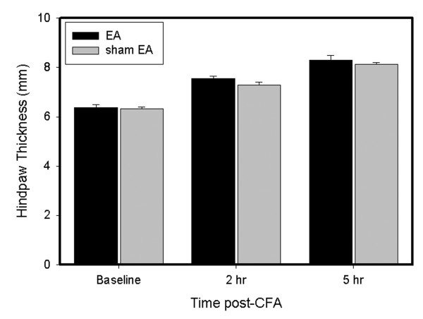
Three experiments were conducted on male Sprague-Dawley rats (n = 7-8/per group). Inflammation was induced by injecting complete Freund's adjuvant (CFA) subcutaneously into the plantar surface of one hind paw. Experiment 1 measured plasma corticosterone (CORT) levels to see if EA regulates CORT secretion. Experiment 2 studied the effects of the adrenal gland on the therapeutic actions of EA using adrenalectomy (ADX) rats. Experiment 3 determined whether a prototypical glucocorticoid receptor antagonist, RU486, affects EA anti-edema. EA treatment, 10 Hz at 3 mA and 0.1 ms pulse width, was given twice, for 20 min each, once immediately after CFA administration and again 2 h post-CFA. Plasma CORT levels, paw thickness, indicative of the intensity of inflammation, and paw withdrawal latency (PWL) were measured 2 h and 5 h after the CFA injection.
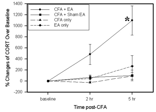
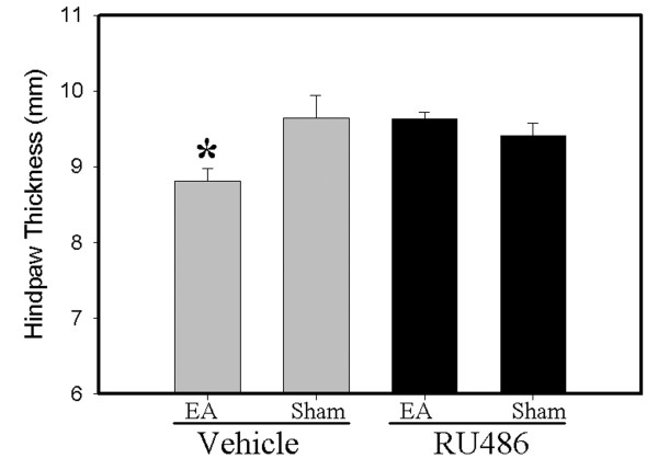
EA significantly increased plasma corticosterone levels 2 h (5 folds) and 5 h (10 folds) after CFA administration compared to sham EA control, but EA alone in naive rats and CFA alone did not induce significant increases in corticosterone. Adrenalectomy blocked EA-produced anti-edema, but not EA anti-hyperalgesia. RU486 (15 mul, 15 mug/mul), a prototypical glucocorticoid receptor antagonist, also prevented EA anti-edema.
The data demonstrate that EA activates the adrenals to increase plasma corticosterone levels and suppress edema and suggest that EA effects differ in healthy subjects and in those with pathologies.
据报道,电针(EA)对炎症性疾病具有抗水肿和抗痛觉过敏作用。然而,其机制尚不清楚。本研究在大鼠模型中探讨了电针抗炎的生化机制。
对雄性Sprague-Dawley大鼠(每组n = 7 - 8只)进行了三项实验。通过将完全弗氏佐剂(CFA)皮下注射到一只后爪的足底表面诱导炎症。实验1测量血浆皮质酮(CORT)水平,以观察电针是否调节CORT分泌。实验2使用肾上腺切除术(ADX)大鼠研究肾上腺对电针治疗作用的影响。实验3确定典型的糖皮质激素受体拮抗剂RU486是否影响电针的抗水肿作用。电针治疗采用3 mA、10 Hz、0.1 ms脉冲宽度,共进行两次,每次20分钟,一次在给予CFA后立即进行,另一次在给予CFA后2小时进行。在注射CFA后2小时和5小时测量血浆CORT水平、爪厚度(指示炎症强度)和爪撤离潜伏期(PWL)。
与假电针对照组相比,电针在给予CFA后2小时(5倍)和5小时(10倍)显著提高了血浆皮质酮水平,但单纯电针处理的未处理大鼠和单纯CFA处理均未引起皮质酮显著升高。肾上腺切除术阻断了电针产生的抗水肿作用,但未阻断电针的抗痛觉过敏作用。典型的糖皮质激素受体拮抗剂RU486(15 μl,15 μg/μl)也可预防电针的抗水肿作用。
数据表明,电针激活肾上腺以增加血浆皮质酮水平并抑制水肿,提示电针在健康受试者和患病受试者中的作用有所不同。