Borsook David, Moulton Eric A, Schmidt Karl F, Becerra Lino R
PAIN Group, Brain Imaging Center, McLean Hospital, 115 Mill Street, Belmont, MA 02478, USA.
Mol Pain. 2007 Sep 11;3:25. doi: 10.1186/1744-8069-3-25.
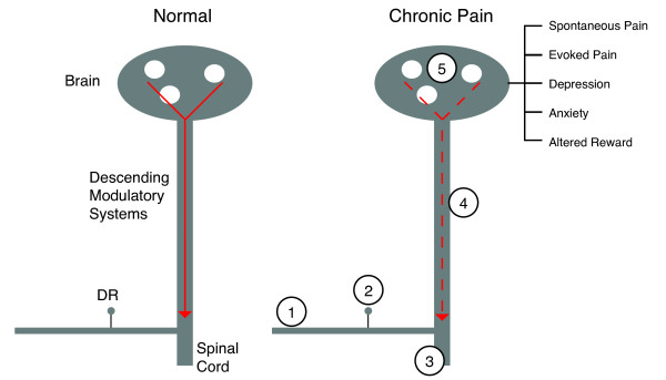
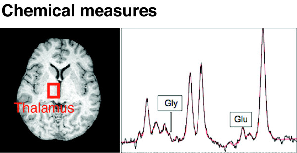
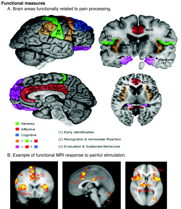
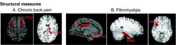
An understanding of how the brain changes in chronic pain or responds to pharmacological or other therapeutic interventions has been significantly changed as a result of developments in neuroimaging of the CNS. These developments have occurred in 3 domains : (1) Anatomical Imaging which has demonstrated changes in brain volume in chronic pain; (2) Functional Imaging (fMRI) that has demonstrated an altered state in the brain in chronic pain conditions including back pain, neuropathic pain, and complex regional pain syndromes. In addition the response of the brain to drugs has provided new insights into how these may modify normal and abnormal circuits (phMRI or pharmacological MRI); (3) Chemical Imaging (Magnetic Resonance Spectroscopy or MRS) has helped our understanding of measures of chemical changes in chronic pain. Taken together these three domains have already changed the way in which we think of pain - it should now be considered an altered brain state in which there may be altered functional connections or systems and a state that has components of degenerative aspects of the CNS.
由于中枢神经系统神经成像技术的发展,我们对大脑在慢性疼痛中如何变化或对药物治疗及其他治疗干预如何反应的理解已发生显著改变。这些发展出现在三个领域:(1)解剖成像,它已证明慢性疼痛中脑容量的变化;(2)功能成像(功能磁共振成像),它已证明在慢性疼痛状况(包括背痛、神经性疼痛和复杂性区域疼痛综合征)下大脑状态的改变。此外,大脑对药物的反应为这些药物如何改变正常和异常回路(药物功能磁共振成像或药理磁共振成像)提供了新的见解;(3)化学成像(磁共振波谱或MRS)有助于我们理解慢性疼痛中化学变化的测量。这三个领域合在一起已经改变了我们对疼痛的看法——现在应将其视为一种大脑状态改变,其中可能存在功能连接或系统的改变,以及一种具有中枢神经系统退行性方面成分的状态。