Simmons Andrea Megela, Horowitz Seth S, Brown Rebecca A
Department of Psychology, Brown University, Providence, RI 02912, USA.
Brain Behav Evol. 2008;71(1):41-53. doi: 10.1159/000108610. Epub 2007 Sep 20.
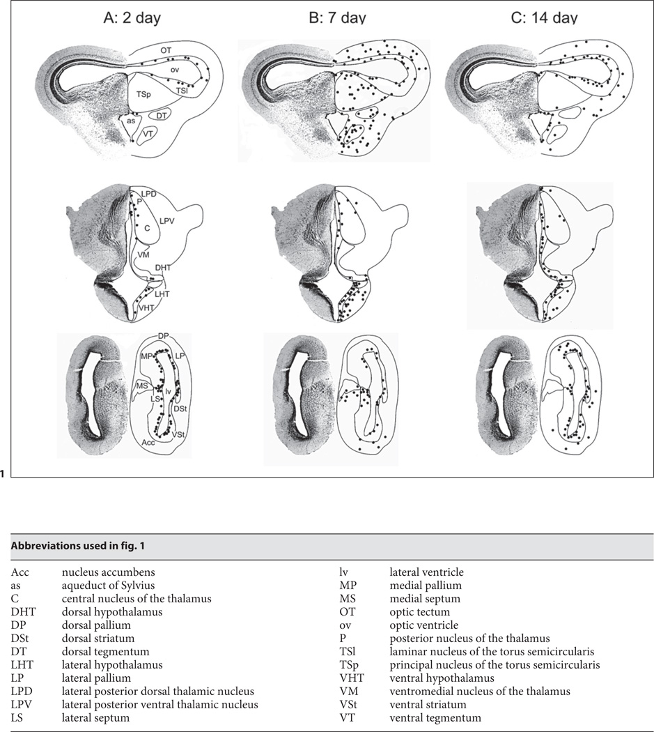
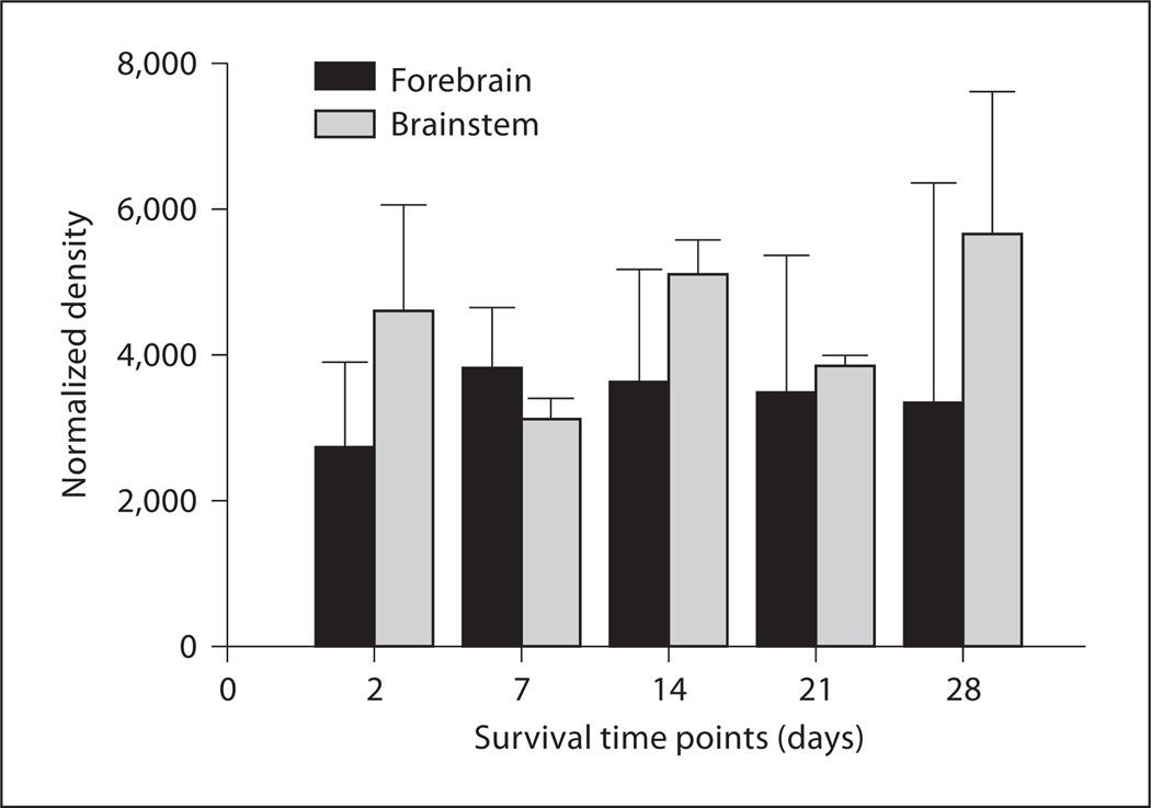
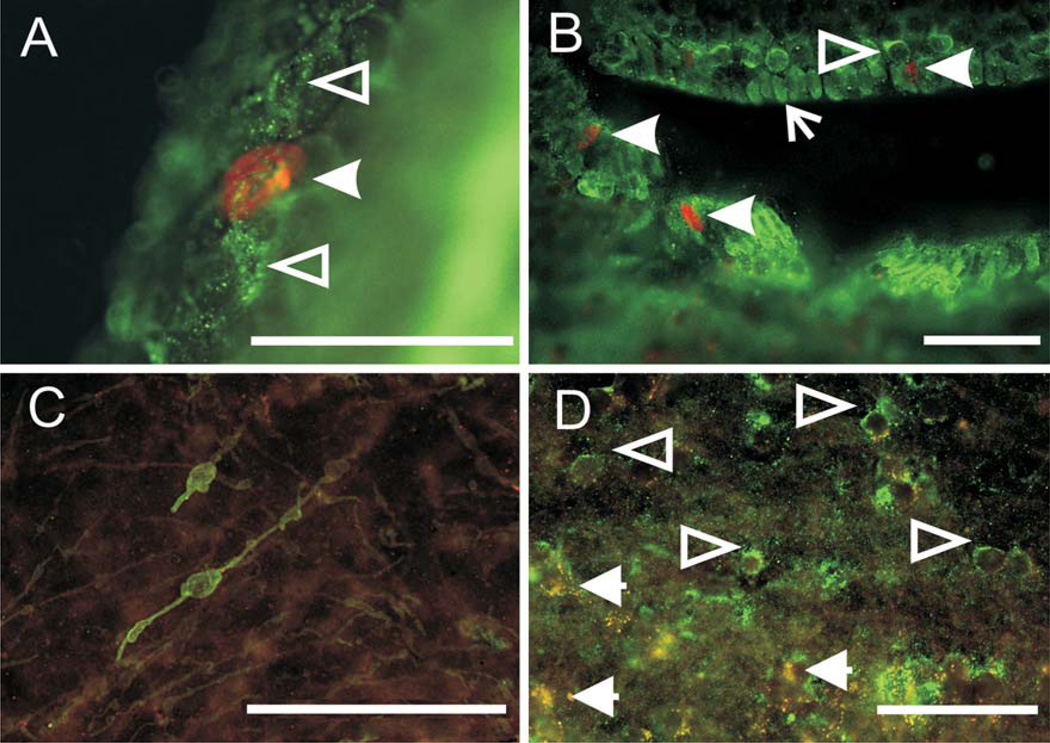
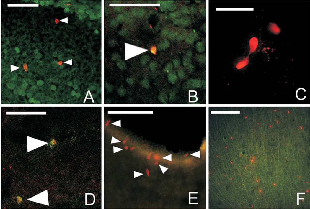
The distribution of proliferating cells in the midbrain, thalamus, and telencephalon of adult bullfrogs (Rana catesbeiana) was examined using immunohistochemistry for the thymidine analog 5-bromo-2'-deoxyuridine (BrdU) and DNA dot-blotting. At all time points examined (2 to 28 days post-injection), BrdU-labeled cells were located in ventricular zones at all levels of the neuraxis, but with relatively more label around the telencephalic ventricles. Labeled cells, some showing profiles indicative of dividing and migrating cells, were present in brain parenchyma from 7 to 28 days post-injection. These labeled cells were particularly numerous in the dorsal and ventral hypothalamus, preoptic area, optic tectum, and laminar and principal nuclei of the torus semicircularis, with label also present, but at qualitatively reduced levels, in thalamic and telencephalic nuclei. Double-label immunohistochemistry using glial and early neural markers indicated that gliogenesis and neurogenesis both occurred, with new neurons observed particularly in the hypothalamus, optic tectum, and torus semicircularis. In all brain areas, many cells not labeled with BrdU were nonetheless labeled with the early neural marker TOAD-64, indicating that these cells were postmitotic. Incorporation of DNA measured by dot-blotting confirms the presence of DNA synthesis in the forebrain and brainstem at all time points measured. The pattern of BrdU label confirms previous experiments based on labeling with (3)H-thymidine and proliferating cell nuclear antigen showing cell proliferation in the adult ranid brain, particularly in hypothalamic nuclei. The consistent appearance of new cells in the hypothalamus of adult frogs suggests that proliferative activity may be important in mediating reproductive behaviors in these animals.
利用针对胸腺嘧啶类似物5-溴-2'-脱氧尿苷(BrdU)的免疫组织化学和DNA斑点印迹法,研究了成年牛蛙(牛蛙)中脑、丘脑和端脑中增殖细胞的分布。在所有检测的时间点(注射后2至28天),BrdU标记的细胞位于神经轴各水平的脑室区,但端脑室周围的标记相对较多。注射后7至28天,脑实质中存在标记细胞,其中一些呈现出分裂和迁移细胞的特征。这些标记细胞在下丘脑背侧和腹侧、视前区、视顶盖以及半规管环层和主核中特别多,丘脑和端脑核中也有标记,但数量在质量上有所减少。使用神经胶质和早期神经标记物的双重标记免疫组织化学表明,神经胶质生成和神经发生都发生了,特别是在下丘脑、视顶盖和半规管环层观察到了新的神经元。在所有脑区,许多未被BrdU标记的细胞却被早期神经标记物TOAD-64标记,这表明这些细胞已完成有丝分裂。通过斑点印迹法测量的DNA掺入证实了在所测量的所有时间点前脑和脑干中都存在DNA合成。BrdU标记模式证实了先前基于用³H-胸腺嘧啶和增殖细胞核抗原标记的实验,显示成年蛙脑中存在细胞增殖,特别是在下丘脑核中。成年蛙下丘脑中持续出现新细胞表明增殖活动可能在介导这些动物的生殖行为中起重要作用。