Lee Meng-Luen
Department of Pediatrics, Division of Pediatric Cardiology, Changhua Christian Hospital, No. 135 Nanhsiao St., Changhua, Taiwan.
Yonsei Med J. 2007 Oct 31;48(5):818-26. doi: 10.3349/ymj.2007.48.5.818.
The clinical and radiological characteristics of the double aortic arch (DAA) and its differentiation from conotruncal malformations (CTM) were reported in order to familiarize pediatric practitioners with these congenital heart diseases.
From July 1994 to December 2006, a total of 6 patients (4 male and 2 female, aged 16 days to 6.5 years) with DAA were enrolled in this retrospective study. The study modalities included chart recordings, plain chest radiographs, barium esophagograms, echocardiograms, cardiac catheterization, cardiac angiograms, surgery, magnetic resonance imaging, and chromosome analysis. Patients with incomplete vascular rings or with right aortic arches and left ligamentum were excluded. In addition, the clinical and radiological profiles of 38 patients with CTM, including dextro-transposition of the great arteries (d-TGA) (n=28), hemitruncus arteriosus (HTA) (n=3), type I truncus arteriosus (TA) (n=4), and the aortopulmonary window (APW) (n=3), were comparatively reviewed.
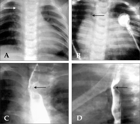
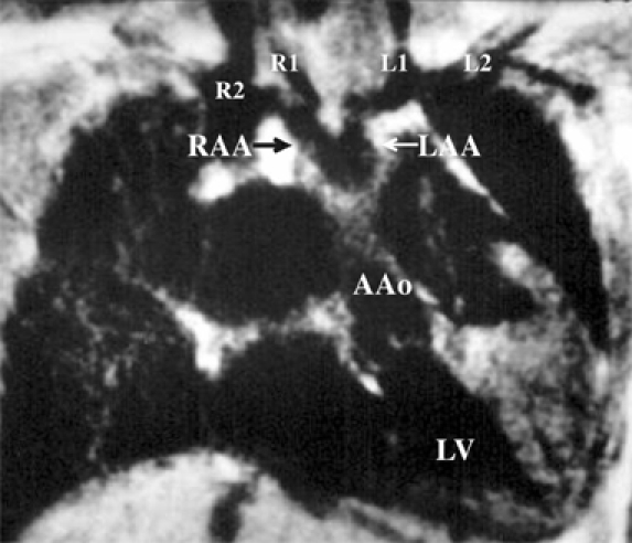
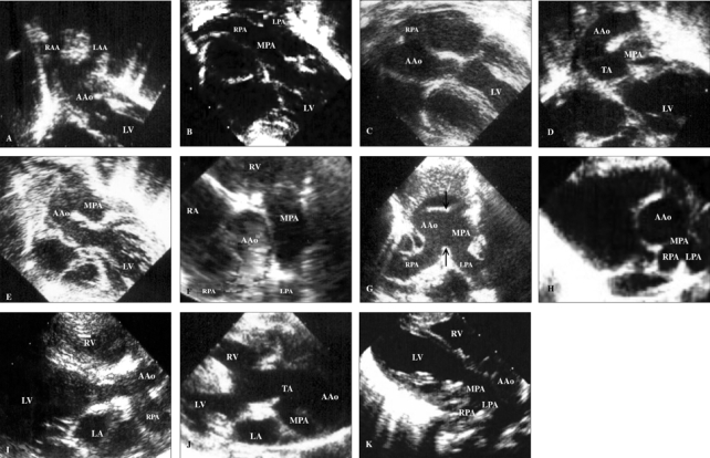
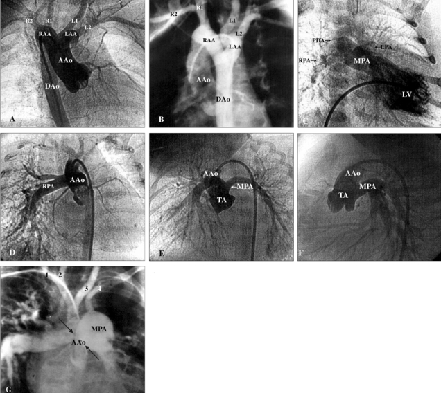
All 6 patients with DAA presented with postprandial choking and respiratory distress that prompted their initial visit to the hospital. One of the 6 patients presented with congestive heart failure, and none with cyanosis. Esophagograms showed indentations in 5 patients with DAA. All patients with d-TGA presented with cyanosis and heart failure, while patients with HTA, type I TA, and APW manifested overt heart failure. Suprasternal and subcostal approaches of the echocardiography may offer diagnostic windows for DAA. As for CTM, parasternal and subcostal approaches could always determine the causality. Cardiac catheterization with angiography comprehensively delineated the pathology.
In case of postprandial choking and respiratory distress in neonates and infants, barium esophagograms can indicate the presence of DAA. Diagnosis of DAA and its differentiation from the CTM can be achieved by echocardiography, angiography, or magnetic resonance imaging.
报告双主动脉弓(DAA)的临床和放射学特征及其与圆锥干畸形(CTM)的鉴别,以使儿科医生熟悉这些先天性心脏病。
1994年7月至2006年12月,共有6例DAA患者(4例男性,2例女性,年龄16天至6.5岁)纳入本回顾性研究。研究方式包括病历记录、胸部X线平片、食管钡餐造影、超声心动图、心导管检查、心血管造影、手术、磁共振成像和染色体分析。排除血管环不完整或右主动脉弓伴左韧带的患者。此外,对38例CTM患者的临床和放射学资料进行了比较性回顾,其中包括大动脉右位转位(d-TGA)(n = 28)、半主动脉干(HTA)(n = 3)、I型主动脉干(TA)(n = 4)和主肺动脉窗(APW)(n = 3)。
6例DAA患者均因餐后呛咳和呼吸窘迫而首次就诊。6例患者中有1例出现充血性心力衰竭,无1例出现紫绀。食管钡餐造影显示5例DAA患者有压迹。所有d-TGA患者均有紫绀和心力衰竭,而HTA、I型TA和APW患者均有明显心力衰竭。超声心动图的胸骨上和肋下途径可为DAA提供诊断窗口。对于CTM,胸骨旁和肋下途径总能确定病因。心导管造影术全面描绘了病变情况。
新生儿和婴儿出现餐后呛咳和呼吸窘迫时,食管钡餐造影可提示DAA的存在。DAA的诊断及其与CTM的鉴别可通过超声心动图、血管造影或磁共振成像来实现。