Petrella Jeffrey R, Prince Steven E, Wang Lihong, Hellegers Caroline, Doraiswamy P Murali
Alzheimer Imaging Research Laboratory and Brain Imaging and Analysis Center, Department of Radiology, Duke University Medical Center, Durham, North Carolina, United States of America.
PLoS One. 2007 Oct 31;2(10):e1104. doi: 10.1371/journal.pone.0001104.
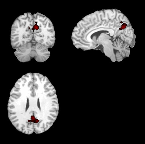
Normal subjects deactivate specific brain regions, notably the posteromedial cortex (PMC), during many tasks. Recent cross-sectional functional magnetic resonance imaging (fMRI) data suggests that deactivation during memory tasks is impaired in Alzheimer's disease (AD). The goal of this study was to prospectively determine the prognostic significance of PMC deactivation in mild cognitive impairment (MCI).
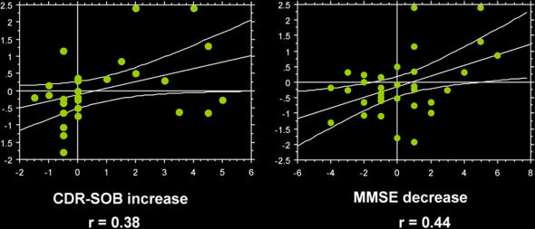
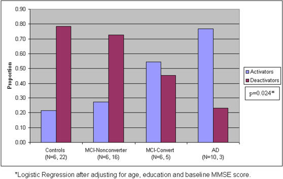
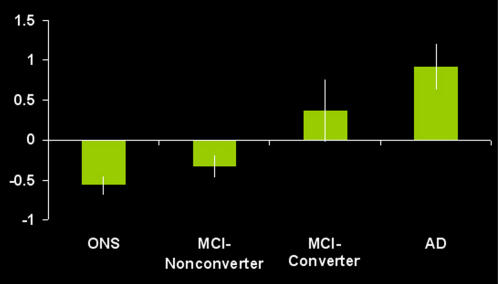
METHODOLOGY/PRINCIPAL FINDINGS: 75 subjects (34 MCI, 13 AD subjects and 28 controls) underwent baseline fMRI scanning during encoding of novel and familiar face-name pairs. MCI subjects were followed longitudinally to determine conversion to AD. Regression and analysis of covariance models were used to assess the effect of PMC activation/deactivation on conversion to dementia as well as in the longitudinal change in dementia measures. At longitudinal follow up of up to 3.5 years (mean 2.5+/-0.79 years), 11 MCI subjects converted to AD. The proportion of deactivators was significantly different across all groups: controls (79%), MCI-Nonconverters (73%), MCI-converters (45%), and AD (23%) (p<0.05). Mean PMC activation magnitude parameter estimates, at baseline, were negative in the control (-0.57+/-0.12) and MCI-Nonconverter (-0.33+/-0.14) groups, and positive in the MCI-Converter (0.37+/-0.40) and AD (0.92+/-0.30) groups. The effect of diagnosis on PMC deactivation remained significant after adjusting for age, education and baseline Mini-Mental State Exam (p<0.05). Baseline PMC activation magnitude was correlated with change in dementia ratings from baseline.
Loss of physiological functional deactivation in the PMC may have prognostic value in preclinical AD, and could aid in profiling subgroups of MCI subjects at greatest risk for progressive cognitive decline.
在许多任务中,正常受试者会使特定脑区失活,尤其是后内侧皮质(PMC)。最近的横断面功能磁共振成像(fMRI)数据表明,阿尔茨海默病(AD)患者在记忆任务期间的失活功能受损。本研究的目的是前瞻性地确定PMC失活在轻度认知障碍(MCI)中的预后意义。
方法/主要发现:75名受试者(34名MCI患者、13名AD患者和28名对照)在对新的和熟悉的面孔-姓名对进行编码期间接受了基线fMRI扫描。对MCI受试者进行纵向随访以确定是否转化为AD。使用回归和协方差分析模型来评估PMC激活/失活对转化为痴呆症的影响以及痴呆症测量指标的纵向变化。在长达3.5年(平均2.5±0.