Park D G, Nam G B, Lee M M, Park Y B, Choi Y S, Seo J D, Lee Y W, Chae H, Kim Y D
Department of Internal Medicine, College of Medicine, Seoul National University, Korea.
Korean J Intern Med. 1991 Jul;6(2):90-8. doi: 10.3904/kjim.1991.6.2.90.
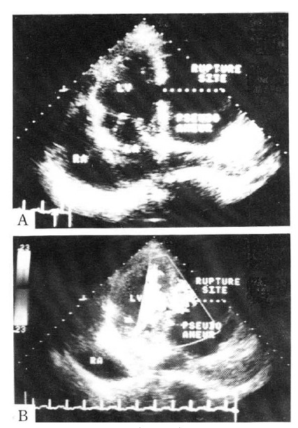
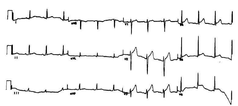
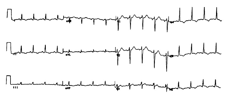
Rupture of the heart as a complication of myocardial infarction is one of the most common causes of in-hospital mortality. Rupture of the free wall of the ventricle or interventricular septum has a poor prognosis when treated conservatively. So, rupture of the heart after infarction requires prompt diagnosis and early surgical repair despite the high overall incidence of early operative mortality before hemodynamic deterioration and multiorgan failures develop. Rupture of the left ventricle results in pseudoaneurysm if the overlying pericardium adhers to the surface of the heart. Pseudoaneurysms which rarely develop after infarction, tend to rupture. Their presence alone is an indicator for operation because of the very poor prognosis following rupture. We experienced successful management of 2 rare complications after acute myocardial infarction: ventricular septal defect and pseudoaneurysm. The first patient was a 49-year-old man who had an apical septal defect. His electrocardiogram showed Q wave in leads V2-V6, II, III, and aVF but a coronary angiogram showed normal findings. He was successfully treated by patch closure of the septal defect. The second patient was a 65-year-old female who had false aneurysm of the left ventricle. She had neither chest pain nor abnormality on the electrocardiogram. A coronary angiogram showed complete occlusion of the distal circumflex artery. Under cardiopulmonary bypass, the neck of the aneurysmal sac was successfully closed with a prolene suture.
心脏破裂作为心肌梗死的并发症是院内死亡的最常见原因之一。心室游离壁或室间隔破裂保守治疗时预后较差。因此,梗死心脏破裂需要及时诊断并尽早进行手术修复,尽管在血流动力学恶化和多器官功能衰竭发生之前早期手术死亡率总体较高。如果覆盖的心包膜粘连于心外膜表面,左心室破裂会导致假性动脉瘤。假性动脉瘤在梗死之后很少发生,且易于破裂。因其破裂后预后极差,仅其存在就是手术指征。我们成功处理了急性心肌梗死后2种罕见并发症:室间隔缺损和假性动脉瘤。首例患者为一名49岁男性,患有心尖部室间隔缺损。其心电图显示V2-V6导联、II、III及aVF导联出现Q波,但冠状动脉造影结果正常。通过补片修补室间隔缺损,他得到了成功治疗。第二例患者是一名65岁女性,患有左心室假性动脉瘤。她既无胸痛,心电图也无异常。冠状动脉造影显示旋支动脉远端完全闭塞。在体外循环下,用普理灵缝线成功闭合了动脉瘤囊颈部。