• 文献检索
  • 文档翻译
  • 深度研究
  • 学术资讯
  • Suppr Zotero 插件Zotero 插件
  • 邀请有礼
  • 套餐&价格
  • 历史记录
应用&插件
Suppr Zotero 插件Zotero 插件浏览器插件Mac 客户端Windows 客户端微信小程序
定价
高级版会员购买积分包购买API积分包
服务
文献检索文档翻译深度研究API 文档MCP 服务
关于我们
关于 Suppr公司介绍联系我们用户协议隐私条款
关注我们

Suppr 超能文献

核心技术专利:CN118964589B侵权必究
粤ICP备2023148730 号-1Suppr @ 2026

文献检索

告别复杂PubMed语法,用中文像聊天一样搜索,搜遍4000万医学文献。AI智能推荐,让科研检索更轻松。

立即免费搜索

文件翻译

保留排版,准确专业,支持PDF/Word/PPT等文件格式,支持 12+语言互译。

免费翻译文档

深度研究

AI帮你快速写综述,25分钟生成高质量综述,智能提取关键信息,辅助科研写作。

立即免费体验

头颈部癌症的治疗后影像学评估。

Post-treatment imaging of head and neck cancer.

机构信息

Department of Radiology, University Hospitals Leuven, Herestraat 49, B-3000 Leuven, Belgium.

出版信息

Cancer Imaging. 2004 Feb 12;4 Spec No A(Spec No A):S6-S15. doi: 10.1102/1470-7330.2004.0007.

DOI:10.1102/1470-7330.2004.0007
PMID:18215976
原文链接:https://pmc.ncbi.nlm.nih.gov/articles/PMC1435341/
Abstract

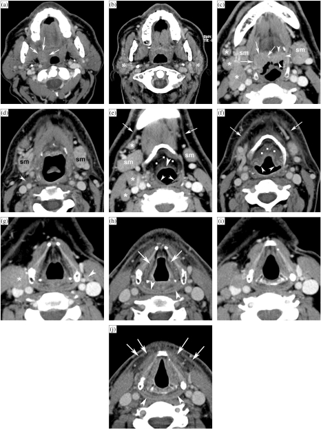
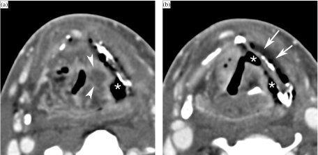
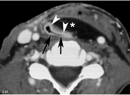
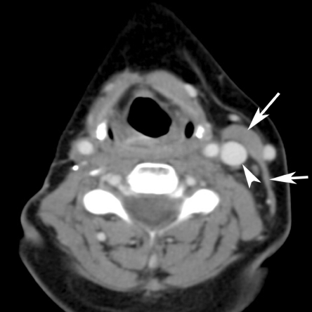
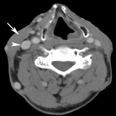
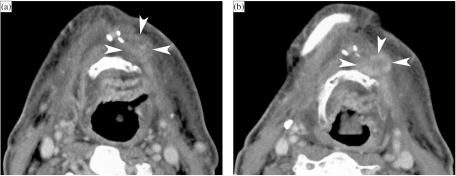
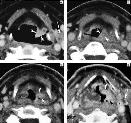
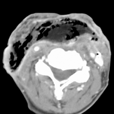
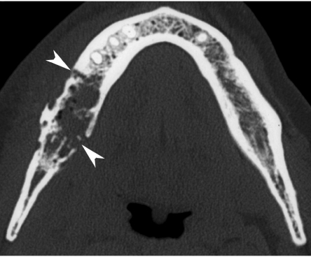
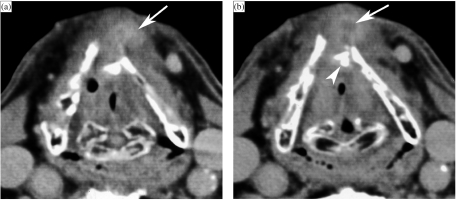
The expected changes on CT or MRI after treatment of a head and neck cancer are described; it is important not to confuse such expected changes with persisting or recurrent tumour, or a treatment complication. Post-treatment CT or MRI is of value when a recurrent tumour is suspected, to confirm the presence of such a lesion and to determine its extent; this is important information for determining the possibility of salvage therapy. More rarely, imaging maybe of use in the differentiation between tumour recurrence and a treatment complication. In patients with a high-risk profile for tumour recurrence after treatment, imaging is of value for surveillance of the patient, as an adjunct to clinical follow-up.The baseline study should be obtained about 3 to 4 months after the end of therapy. There is evidence that tumour recurrences can be detect earlier by systematic follow-up imaging.

摘要

治疗头颈部癌症后 CT 或 MRI 的预期变化;重要的是不要将这些预期变化与持续存在或复发的肿瘤或治疗并发症相混淆。当怀疑复发肿瘤时,治疗后的 CT 或 MRI 具有价值,可以确认存在这种病变并确定其范围;这对于确定挽救治疗的可能性很重要。在极少数情况下,影像学检查可能有助于区分肿瘤复发和治疗并发症。在治疗后肿瘤复发风险高的患者中,影像学检查对患者的监测具有价值,可作为临床随访的辅助手段。基线研究应在治疗结束后约 3 至 4 个月进行。有证据表明,系统的随访影像学检查可以更早地检测到肿瘤复发。

https://cdn.ncbi.nlm.nih.gov/pmc/blobs/8faa/1435341/018749ede969/ci040006.jpg
https://cdn.ncbi.nlm.nih.gov/pmc/blobs/8faa/1435341/051b113529bd/ci04000601.jpg
https://cdn.ncbi.nlm.nih.gov/pmc/blobs/8faa/1435341/78482209405c/ci04000602.jpg
https://cdn.ncbi.nlm.nih.gov/pmc/blobs/8faa/1435341/df7b155175c5/ci04000603.jpg
https://cdn.ncbi.nlm.nih.gov/pmc/blobs/8faa/1435341/2118ff8f5f5f/ci04000604.jpg
https://cdn.ncbi.nlm.nih.gov/pmc/blobs/8faa/1435341/dbdf5a050719/ci04000605.jpg
https://cdn.ncbi.nlm.nih.gov/pmc/blobs/8faa/1435341/96224e44bd63/ci04000606.jpg
https://cdn.ncbi.nlm.nih.gov/pmc/blobs/8faa/1435341/e8d6d6a51f22/ci04000607.jpg
https://cdn.ncbi.nlm.nih.gov/pmc/blobs/8faa/1435341/b09c85e43d0d/ci04000608.jpg
https://cdn.ncbi.nlm.nih.gov/pmc/blobs/8faa/1435341/5d209bcf3bbf/ci04000609.jpg
https://cdn.ncbi.nlm.nih.gov/pmc/blobs/8faa/1435341/018749ede969/ci040006.jpg

相似文献

1
Post-treatment imaging of head and neck cancer.头颈部癌症的治疗后影像学评估。
Cancer Imaging. 2004 Feb 12;4 Spec No A(Spec No A):S6-S15. doi: 10.1102/1470-7330.2004.0007.
2
Posttreatment imaging in head and neck cancer.头颈部癌的治疗后影像学检查
Eur J Radiol. 2008 Jun;66(3):501-11. doi: 10.1016/j.ejrad.2008.01.021. Epub 2008 Mar 6.
3
FDG-PET delayed imaging for the detection of head and neck cancer recurrence after radio-chemotherapy: comparison with MRI/CT.氟代脱氧葡萄糖正电子发射断层显像(FDG-PET)延迟显像用于检测放化疗后头颈癌复发:与磁共振成像(MRI)/计算机断层扫描(CT)的比较
Eur J Nucl Med Mol Imaging. 2004 Apr;31(4):590-5. doi: 10.1007/s00259-003-1408-6. Epub 2004 Jan 14.
4
Imaging modalities in head-and-neck cancer patients.头颈部癌症患者的成像模态
Indian J Dent Res. 2012 Nov-Dec;23(6):819-21. doi: 10.4103/0970-9290.111270.
5
Posttreatment Imaging in Patients with Head and Neck Cancer without Clinical Evidence of Recurrence: Should Surveillance Imaging Extend Beyond 6 Months?头颈部癌症患者治疗后无临床复发证据的影像学检查:监测影像学检查是否应延长至 6 个月以上?
AJNR Am J Neuroradiol. 2020 Jul;41(7):1238-1244. doi: 10.3174/ajnr.A6614. Epub 2020 Jun 18.
6
Locoregional tumour evaluation of squamous cell carcinoma in the head and neck area: a comparison between MRI, PET/CT and integrated PET/MRI.头颈部鳞状细胞癌的局部区域肿瘤评估:MRI、PET/CT与PET/MRI一体化成像的比较
Eur J Nucl Med Mol Imaging. 2016 Jan;43(1):92-102. doi: 10.1007/s00259-015-3145-z. Epub 2015 Aug 6.
7
Local recurrence of squamous cell carcinoma of the head and neck after radio(chemo)therapy: Diagnostic performance of FDG-PET/MRI with diffusion-weighted sequences.头颈部鳞状细胞癌放(化)疗后局部复发:FDG-PET/MRI 联合弥散加权序列的诊断性能。
Eur Radiol. 2018 Feb;28(2):651-663. doi: 10.1007/s00330-017-4999-1. Epub 2017 Aug 15.
8
F-FDG PET/CT surveillance for the detection of recurrence in patients with head and neck cancer.用于检测头颈癌患者复发情况的F-FDG PET/CT监测
Eur J Cancer. 2017 Feb;72:62-70. doi: 10.1016/j.ejca.2016.11.009. Epub 2016 Dec 24.
9
Effect of (18)F-fluorodeoxyglucose positron emission tomography/computed tomography-guided management of suspected recurrent papillary thyroid carcinoma: long-term follow-up with tumour marker responses.(18)F-氟代脱氧葡萄糖正电子发射断层扫描/计算机断层扫描引导下对疑似复发性甲状腺乳头状癌的管理:肿瘤标志物反应的长期随访。
Clin Oncol (R Coll Radiol). 2012 Dec;24(10):e168-72. doi: 10.1016/j.clon.2012.08.002. Epub 2012 Sep 1.
10
Recurrent head and neck cancer: United Kingdom National Multidisciplinary Guidelines.复发性头颈癌:英国国家多学科指南
J Laryngol Otol. 2016 May;130(S2):S181-S190. doi: 10.1017/S002221511600061X.

引用本文的文献

1
Comprehensive review of post-treatment imaging in head and neck cancers: from expected to unexpected and beyond.头颈部癌症治疗后影像学的全面综述:从预期到意外及其他。
Br J Radiol. 2024 Dec 1;97(1164):1898-1914. doi: 10.1093/bjr/tqae207.
2
Quantitative measurements of radiation-induced fibrosis for head and neck cancer: A narrative review.头颈部癌放疗后纤维化的定量测量:一篇叙述性综述
Laryngoscope Investig Otolaryngol. 2024 Apr 22;9(2):e1249. doi: 10.1002/lio2.1249. eCollection 2024 Apr.
3
Postoperative Imaging Appearance of Iliac Crest Free Flaps Used for Palatomaxillary Reconstructions.

本文引用的文献

1
Postradiotherapy surveillance practice for head and neck squamous cell carcinoma--too much for too little?头颈部鳞状细胞癌放疗后的监测实践——是否做得过多而收获过少?
Head Neck. 2003 Dec;25(12):990-9. doi: 10.1002/hed.10314.
2
Imaging of mandibular osteoradionecrosis.
Neuroimaging Clin N Am. 2003 Aug;13(3):597-604. doi: 10.1016/s1052-5149(03)00036-4.
3
Evaluation of head and neck squamous cell carcinoma after treatment.治疗后头颈部鳞状细胞癌的评估
AJNR Am J Neuroradiol. 2003 Oct;24(9):1743-6.
用于腭上颌重建的髂嵴游离皮瓣的术后影像学表现。
AJNR Am J Neuroradiol. 2021 Apr;42(4):753-758. doi: 10.3174/ajnr.A7005. Epub 2021 Feb 25.
4
Comparison of tumor delineation using dual energy computed tomography versus magnetic resonance imaging in head and neck cancer re-irradiation cases.头颈部癌再程放疗病例中使用双能计算机断层扫描与磁共振成像进行肿瘤勾画的比较。
Phys Imaging Radiat Oncol. 2020 Apr 29;14:1-5. doi: 10.1016/j.phro.2020.04.001. eCollection 2020 Apr.
5
Early Assessment of Tumor Response to Radiation Therapy using High-Resolution Quantitative Microvascular Ultrasound Imaging.使用高分辨率定量微血管超声成像技术早期评估肿瘤对放射治疗的反应。
Theranostics. 2018 Jan 1;8(1):156-168. doi: 10.7150/thno.19703. eCollection 2018.
6
Role of imaging in the follow-up of T2-T3 glottic cancer treated by transoral laser microsurgery.影像学在经口激光显微手术治疗T2-T3声门癌随访中的作用
Eur Arch Otorhinolaryngol. 2017 Oct;274(10):3679-3686. doi: 10.1007/s00405-017-4642-4. Epub 2017 Jun 19.
7
Postoperative Imaging and Surveillance in Large Nerve Perineural Spread.大型神经周围扩散的术后影像学检查与监测
J Neurol Surg B Skull Base. 2016 Apr;77(2):182-92. doi: 10.1055/s-0036-1571840. Epub 2016 Feb 9.
8
Feasibility analysis of the parametric response map as an early predictor of treatment efficacy in head and neck cancer.参数反应图作为头颈癌治疗疗效早期预测指标的可行性分析
AJNR Am J Neuroradiol. 2015 Apr;36(4):757-62. doi: 10.3174/ajnr.A4296. Epub 2015 Mar 19.
9
Which is the most reliable diagnostic modality for detecting locally residual or recurrent laryngeal squamous cell carcinoma after (chemo)radiotherapy?对于检测(化疗)放疗后局部残留或复发性喉鳞状细胞癌,哪种诊断方式最可靠?
Eur Arch Otorhinolaryngol. 2013 Nov;270(11):2787-91. doi: 10.1007/s00405-013-2564-3. Epub 2013 May 21.
10
Limited utility of routine surveillance MRI following chemoradiation for advanced-stage oropharynx carcinoma.同步放化疗后常规监测性磁共振成像在晚期口咽癌中的应用价值有限。
Int J Otolaryngol. 2010;2010. doi: 10.1155/2010/904297. Epub 2010 Aug 31.
4
MICROPATHOLOGY OF RADIATION REACTION IN THE LARYNX.喉部放射反应的微观病理学
Ann Otol Rhinol Laryngol. 1963 Sep;72:831-41. doi: 10.1177/000348946307200316.
5
MRI appearance of radiation-induced changes of normal cervical tissues.正常宫颈组织辐射诱导变化的磁共振成像表现
Eur Radiol. 2001;11(9):1807-17. doi: 10.1007/s003300000728.
6
CT findings in laryngeal chondroradionecrosis.喉放射性软骨坏死的CT表现
JBR-BTR. 1999 Feb;82(1):16-8.
7
Laryngeal or hypopharyngeal squamous cell carcinoma: can follow-up CT after definitive radiation therapy be used to detect local failure earlier than clinical examination alone?喉或下咽鳞状细胞癌:在根治性放射治疗后,随访CT能否比单纯临床检查更早地检测出局部复发?
Radiology. 2000 Mar;214(3):683-7. doi: 10.1148/radiology.214.3.r00fe13683.
8
Masticator space abnormalities associated with mandibular osteoradionecrosis: MR and CT findings in five patients.与下颌骨放射性骨坏死相关的咀嚼肌间隙异常:5例患者的磁共振成像(MR)和计算机断层扫描(CT)表现
AJNR Am J Neuroradiol. 2000 Jan;21(1):175-8.
9
Pre- and post-radiotherapy computed tomography in laryngeal cancer: imaging-based prediction of local failure.喉癌放疗前后的计算机断层扫描:基于影像学的局部失败预测
Int J Radiat Oncol Biol Phys. 1999 Sep 1;45(2):359-66. doi: 10.1016/s0360-3016(99)00149-2.
10
An imaging-based classification for the cervical nodes designed as an adjunct to recent clinically based nodal classifications.一种基于影像学的颈部淋巴结分类方法,旨在作为近期基于临床的淋巴结分类的辅助手段。
Arch Otolaryngol Head Neck Surg. 1999 Apr;125(4):388-96. doi: 10.1001/archotol.125.4.388.