UNC/NCSU Joint Dept. of Biomedical Engineering.
Sonovol, Inc. (current affiliation).
Theranostics. 2018 Jan 1;8(1):156-168. doi: 10.7150/thno.19703. eCollection 2018.
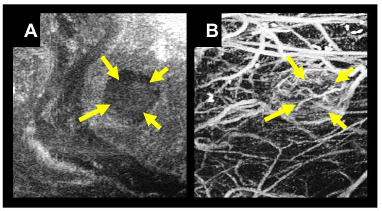
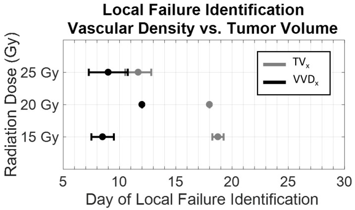
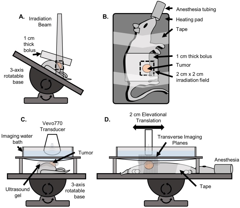
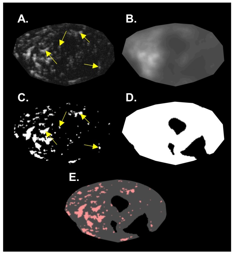
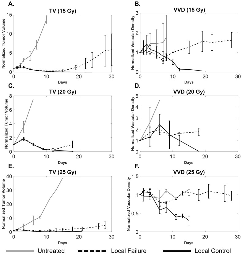
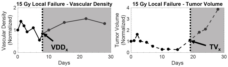
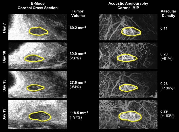
Measuring changes in tumor volume using anatomical imaging weeks to months post radiation therapy (RT) is currently the clinical standard for indicating treatment response to RT. For patients whose tumors do not respond successfully to treatment, this approach is suboptimal as timely modification of the treatment approach may lead to better clinical outcomes. We propose to use tumor microvasculature as a biomarker for early assessment of tumor response to RT. Acoustic angiography is a novel contrast ultrasound imaging technique that enables high-resolution microvascular imaging and has been shown to detect changes in microvascular structure due to cancer growth. Data suggest that acoustic angiography can detect longitudinal changes in the tumor microvascular environment that correlate with RT response. Three cohorts of Fisher 344 rats were implanted with rat fibrosarcoma tumors and were treated with a single fraction of RT at three dose levels (15 Gy, 20 Gy, and 25 Gy) at a dose rate of 300 MU/min. A simple treatment condition was chosen for testing the feasibility of our imaging technique. All tumors were longitudinally imaged immediately prior to and after treatment and then every 3 days after treatment for a total of 30 days. Both acoustic angiography (using in-house produced microbubble contrast agents) and standard b-mode imaging was performed at each imaging time point using a pre-clinical Vevo770 scanner and a custom modified dual-frequency transducer. Results show that all treated tumors in each dose group initially responded to treatment between days 3-15 as indicated by decreased tumor growth accompanied with decreased vascular density. Untreated tumors continued to increase in both volume and vascular density until they reached the maximum allowable size of 2 cm in diameter. Tumors that displayed complete control (no tumor recurrence) continued to decrease in size and vascular density, while tumors that progressed after the initial response presented an increase in tumor volume and volumetric vascular density. The increase in tumor volumetric vascular density in recurring tumors can be detected 10.25 ± 1.5 days, 6 ± 0 days, and 4 ± 1.4 days earlier than the measurable increase in tumor volume in the 15, 20, and 25 Gy dose groups, respectively. A dose-dependent growth rate for tumor recurrence was also observed. In this feasibility study we have demonstrated the ability of acoustic angiography to detect longitudinal changes in vascular density, which was shown to be a potential biomarker for tumor response to RT.
使用解剖成像技术在放射治疗 (RT) 后数周到数月测量肿瘤体积的变化,目前是临床判断 RT 治疗反应的标准。对于肿瘤对治疗反应不成功的患者,这种方法并不理想,因为及时修改治疗方法可能会带来更好的临床结果。我们建议使用肿瘤微血管作为早期评估肿瘤对 RT 反应的生物标志物。声振血管造影是一种新的对比超声成像技术,可实现高分辨率微血管成像,并已被证明可检测到由于癌症生长导致的微血管结构变化。数据表明,声振血管造影可以检测到与 RT 反应相关的肿瘤微血管环境的纵向变化。三个 Fisher 344 大鼠队列被植入大鼠纤维肉瘤肿瘤,并在三种剂量水平(15 Gy、20 Gy 和 25 Gy)下接受单次 RT 治疗,剂量率为 300 MU/min。选择了一种简单的治疗条件来测试我们的成像技术的可行性。所有肿瘤在治疗前和治疗后立即进行纵向成像,然后在治疗后每 3 天进行一次成像,总共 30 天。在每个成像时间点,使用临床前 Vevo770 扫描仪和定制的双频换能器进行声振血管造影(使用内部生产的微泡造影剂)和标准 B 模式成像。结果表明,在每个剂量组中,所有接受治疗的肿瘤最初在第 3-15 天内对治疗有反应,表现为肿瘤生长减少并伴有血管密度降低。未接受治疗的肿瘤在体积和血管密度方面继续增加,直到达到直径 2 厘米的最大允许尺寸。显示完全控制(无肿瘤复发)的肿瘤继续减小体积和血管密度,而在初始反应后进展的肿瘤则表现出肿瘤体积和体积血管密度的增加。复发肿瘤的肿瘤体积血管密度增加可分别提前 10.25 ± 1.5 天、6 ± 0 天和 4 ± 1.4 天检测到,而在 15、20 和 25 Gy 剂量组中,可检测到肿瘤体积的可测量增加。还观察到肿瘤复发的剂量依赖性生长率。在这项可行性研究中,我们已经证明了声振血管造影检测血管密度纵向变化的能力,这表明它是肿瘤对 RT 反应的潜在生物标志物。