Winkeler A, Waerzeggers Y, Klose A, Monfared P, Thomas A V, Schubert M, Heneka M T, Jacobs A H
Laboratory for Gene Therapy and Molecular Imaging, Max Planck Institute for Neurological Research, and the Faculty of Medicine, University of Cologne, Gleuelerstrasse 50, Cologne, Germany.
Eur J Nucl Med Mol Imaging. 2008 Mar;35 Suppl 1(Suppl 1):S107-13. doi: 10.1007/s00259-007-0710-0.
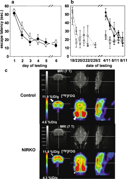
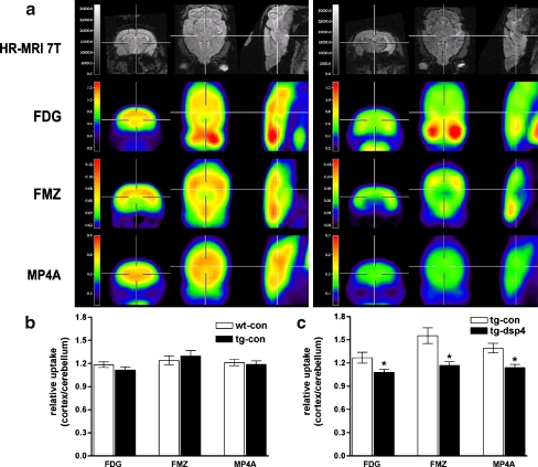
Molecular imaging aims towards the non-invasive characterization of disease-specific molecular alterations in the living organism in vivo. In that, molecular imaging opens a new dimension in our understanding of disease pathogenesis, as it allows the non-invasive determination of the dynamics of changes on the molecular level. IMAGING OF AD CHARACTERISTIC CHANGES BY microPET: The imaging technology being employed includes magnetic resonance imaging (MRI) and nuclear imaging as well as optical-based imaging technologies. These imaging modalities are employed together or alone for disease phenotyping, development of imaging-guided therapeutic strategies and in basic and translational research. In this study, we review recent investigations employing positron emission tomography and MRI for phenotyping mouse models of Alzheimer's disease by imaging. We demonstrate that imaging has an important role in the characterization of mouse models of neurodegenerative diseases.
分子成像旨在对活体生物体内疾病特异性分子改变进行非侵入性表征。就此而言,分子成像为我们理解疾病发病机制开启了一个新维度,因为它能够非侵入性地测定分子水平变化的动态过程。通过微型正电子发射断层扫描(microPET)对阿尔茨海默病特征性变化进行成像:所采用的成像技术包括磁共振成像(MRI)、核成像以及基于光学的成像技术。这些成像方式可单独或联合用于疾病表型分析、成像引导治疗策略的开发以及基础和转化研究。在本研究中,我们回顾了近期通过成像利用正电子发射断层扫描和MRI对阿尔茨海默病小鼠模型进行表型分析的研究。我们证明成像在神经退行性疾病小鼠模型的表征中具有重要作用。