Chemical Biology Program, Memorial Sloan Kettering Cancer Center, New York, NY, 10065, USA.
Program of Neuroscience, Weill Graduate School of Medical Sciences of Cornell University, New York, NY, 10021, USA.
Sci Rep. 2020 Jun 25;10(1):10379. doi: 10.1038/s41598-020-67284-z.
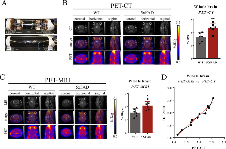
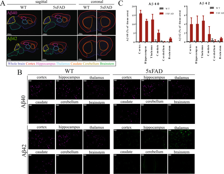
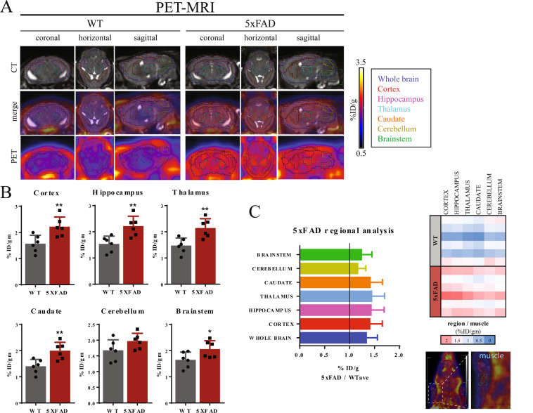
The emergence of PET probes for amyloid plaques and neurofibrillary tangles, hallmarks of Alzheimer disease (AD), enables monitoring of pathology in AD mouse models. However, small-animal PET imaging is limited by coarse spatial resolution. We have installed a custom-fabricated PET insert into our small-animal MRI instrument and used PET/MRI hybrid imaging to define regions of amyloid vulnerability in 5xFAD mice. We compared fluorine-18 [F]-Florbetapir uptake in the 5xFAD brain by dedicated small-animal PET/MRI and PET/CT to validate the quantitative measurement of PET/MRI. Next, we used PET/MRI to define uptake in six brain regions. As expected, uptake was comparable to wild-type in the cerebellum and elevated in the cortex and hippocampus, regions implicated in AD. Interestingly, uptake was highest in the thalamus, a region often overlooked in AD studies. Development of small-animal PET/MRI enables tracking of brain region-specific pathology in mouse models, which may prove invaluable to understanding AD progression and therapeutic development.
淀粉样斑块和神经原纤维缠结的 PET 探针的出现,是阿尔茨海默病(AD)的标志,能够监测 AD 小鼠模型中的病理学。然而,小动物 PET 成像受到粗糙空间分辨率的限制。我们已经在我们的小动物 MRI 仪器中安装了定制的 PET 插件,并使用 PET/MRI 混合成像来定义 5xFAD 小鼠中的淀粉样脆弱区域。我们通过专用的小动物 PET/MRI 和 PET/CT 比较了 5xFAD 大脑中的氟-18 [F]-Florbetapir 摄取,以验证 PET/MRI 的定量测量。接下来,我们使用 PET/MRI 来定义六个脑区的摄取。正如预期的那样,小脑的摄取与野生型相似,而大脑皮层和海马体的摄取升高,这些区域与 AD 有关。有趣的是,摄取在丘脑最高,这是 AD 研究中经常被忽视的区域。小动物 PET/MRI 的发展使得能够在小鼠模型中追踪特定脑区的病理学,这对于理解 AD 进展和治疗开发可能非常有价值。