Hui David Y
Department of Pathology and Laboratory Medicine, Genome Research Institute, University of Cincinnati College of Medicine, Cincinnati, Ohio 45237, USA.
Curr Drug Targets. 2008 Mar;9(3):251-60. doi: 10.2174/138945008783755601.
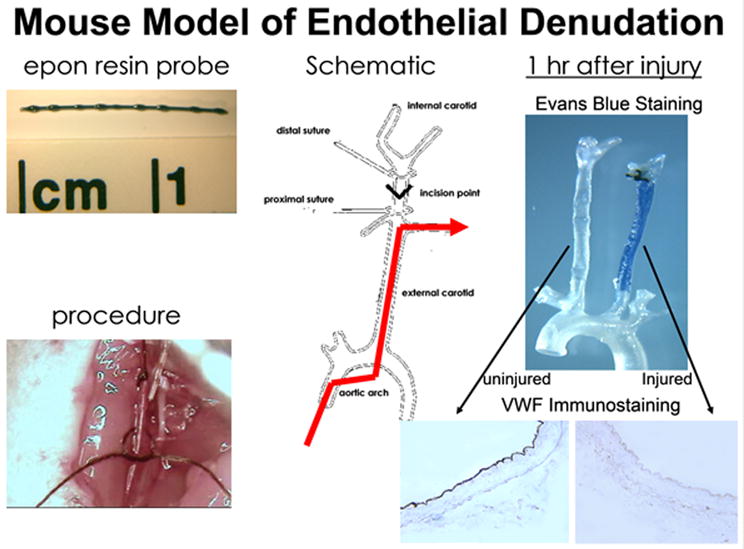
The most commonly used procedures to induce arterial injury in mice are carotid artery ligation with cessation of blood flow and mechanically-induced denudation of endothelium in the carotid or the femoral arteries. Both procedures result in neointimal hyperplasia after two to three weeks. A survey of various inbred strains of mice shows that strain-specific differences in susceptibility to injury-induced neointimal hyperplasia are different than those for susceptibility to diet-induced atherosclerosis, with strains identified as susceptible to both neointimal hyperplasia and atherosclerosis, resistant to both, susceptible to atherosclerosis but resistant to neointimal hyperplasia, or resistant to atherosclerosis but susceptible to neointimal hyperplasia. Inflammatory cells such as T and B lymphocytes, which are contributory to atherosclerosis, are protective against injury-induced neointimal hyperplasia. In contrast, the infiltration of monocytes into the site of injury and their differentiation to macrophages favor neointimal hyperplasia similar to their pathogenic role in atherosclerosis. The regulatory role of lymphocytes and macrophages in neointimal hyperplasia is related to the production of cytokines such as interferon-gamma and tumor necrosis factor-alpha, respectively. Interestingly, inducible nitric oxide synthase (iNOS) activity appears to inhibit neointimal hyperplasia in the endothelial denudation model but contributes to neointimal hyperplasia when arterial injury is induced by periadventitial cuff placement. The difference appears to be due to the time required for endothelial recovery and the participation of inflammatory cells. Thus, although arterial injury-induced neointimal hyperplasia results in similar vascular occlusion as progressive atherosclerosis, the pathology and mechanism of the two disease processes are quite different.
在小鼠中诱导动脉损伤最常用的方法是结扎颈动脉并阻断血流,以及机械性剥脱颈动脉或股动脉的内皮。这两种方法在两到三周后都会导致内膜增生。对各种近交系小鼠的调查显示,损伤诱导的内膜增生易感性的品系特异性差异与饮食诱导的动脉粥样硬化易感性不同,已鉴定出对内膜增生和动脉粥样硬化均易感、对两者均有抗性、对动脉粥样硬化易感但对内膜增生有抗性,或对动脉粥样硬化有抗性但对内膜增生易感的品系。促成动脉粥样硬化的T淋巴细胞和B淋巴细胞等炎症细胞对损伤诱导的内膜增生具有保护作用。相反,单核细胞浸润到损伤部位并分化为巨噬细胞有利于内膜增生,类似于它们在动脉粥样硬化中的致病作用。淋巴细胞和巨噬细胞在内膜增生中的调节作用分别与干扰素-γ和肿瘤坏死因子-α等细胞因子的产生有关。有趣的是,诱导型一氧化氮合酶(iNOS)活性在血管内皮剥脱模型中似乎抑制内膜增生,但在通过外膜袖带放置诱导动脉损伤时则促成内膜增生。这种差异似乎是由于内皮恢复所需的时间和炎症细胞的参与。因此,尽管动脉损伤诱导的内膜增生与进行性动脉粥样硬化导致相似的血管闭塞,但这两个疾病过程的病理和机制却大不相同。