• 文献检索
  • 文档翻译
  • 深度研究
  • 学术资讯
  • Suppr Zotero 插件Zotero 插件
  • 邀请有礼
  • 套餐&价格
  • 历史记录
应用&插件
Suppr Zotero 插件Zotero 插件浏览器插件Mac 客户端Windows 客户端微信小程序
定价
高级版会员购买积分包购买API积分包
服务
文献检索文档翻译深度研究API 文档MCP 服务
关于我们
关于 Suppr公司介绍联系我们用户协议隐私条款
关注我们

Suppr 超能文献

核心技术专利:CN118964589B侵权必究
粤ICP备2023148730 号-1Suppr @ 2026

文献检索

告别复杂PubMed语法,用中文像聊天一样搜索,搜遍4000万医学文献。AI智能推荐,让科研检索更轻松。

立即免费搜索

文件翻译

保留排版,准确专业,支持PDF/Word/PPT等文件格式,支持 12+语言互译。

免费翻译文档

深度研究

AI帮你快速写综述,25分钟生成高质量综述,智能提取关键信息,辅助科研写作。

立即免费体验

背景体部信号抑制的扩散加权全身成像(DWIBS):肿瘤学中的特征及潜在应用

Diffusion-weighted whole-body imaging with background body signal suppression (DWIBS): features and potential applications in oncology.

作者信息

Kwee Thomas C, Takahara Taro, Ochiai Reiji, Nievelstein Rutger A J, Luijten Peter R

机构信息

Department of Radiology (HP E01.132), University Medical Center Utrecht, Heidelberglaan 100, 3584 CX, Utrecht, The Netherlands.

出版信息

Eur Radiol. 2008 Sep;18(9):1937-52. doi: 10.1007/s00330-008-0968-z. Epub 2008 Apr 30.

DOI:10.1007/s00330-008-0968-z
PMID:18446344
原文链接:https://pmc.ncbi.nlm.nih.gov/articles/PMC2516183/
Abstract

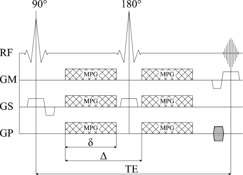
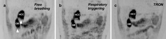
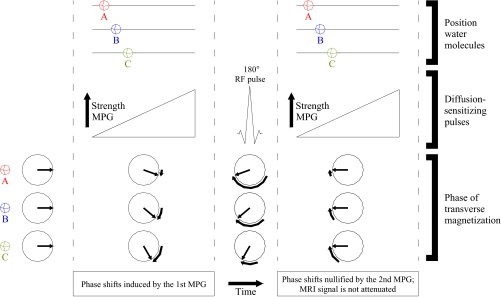
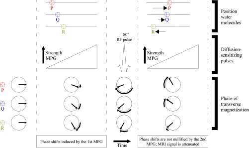
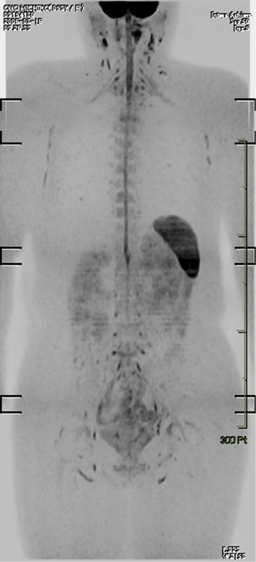
Diffusion-weighted magnetic resonance imaging (DWI) provides functional information and can be used for the detection and characterization of pathologic processes, including malignant tumors. The recently introduced concept of "diffusion-weighted whole-body imaging with background body signal suppression" (DWIBS) now allows acquisition of volumetric diffusion-weighted images of the entire body. This new concept has unique features different from conventional DWI and may play an important role in whole-body oncological imaging. This review describes and illustrates the basics of DWI, the features of DWIBS, and its potential applications in oncology.

摘要

扩散加权磁共振成像(DWI)可提供功能信息,可用于检测和表征包括恶性肿瘤在内的病理过程。最近引入的“背景体素信号抑制扩散加权全身成像”(DWIBS)概念,现在可以获取全身的容积扩散加权图像。这一新概念具有与传统DWI不同的独特特征,可能在全身肿瘤成像中发挥重要作用。本综述描述并阐述了DWI的基础、DWIBS的特征及其在肿瘤学中的潜在应用。

https://cdn.ncbi.nlm.nih.gov/pmc/blobs/a8c4/2516183/891d029b8273/330_2008_968_Fig13_HTML.jpg
https://cdn.ncbi.nlm.nih.gov/pmc/blobs/a8c4/2516183/1f0b7061b766/330_2008_968_Fig1_HTML.jpg
https://cdn.ncbi.nlm.nih.gov/pmc/blobs/a8c4/2516183/0d5e76555b04/330_2008_968_Fig2_HTML.jpg
https://cdn.ncbi.nlm.nih.gov/pmc/blobs/a8c4/2516183/b873fc6d44d5/330_2008_968_Fig3_HTML.jpg
https://cdn.ncbi.nlm.nih.gov/pmc/blobs/a8c4/2516183/3e4bc141d0d1/330_2008_968_Fig4_HTML.jpg
https://cdn.ncbi.nlm.nih.gov/pmc/blobs/a8c4/2516183/76023e9f187d/330_2008_968_Fig5_HTML.jpg
https://cdn.ncbi.nlm.nih.gov/pmc/blobs/a8c4/2516183/8afd704715d1/330_2008_968_Fig6_HTML.jpg
https://cdn.ncbi.nlm.nih.gov/pmc/blobs/a8c4/2516183/51ed8805243d/330_2008_968_Fig7_HTML.jpg
https://cdn.ncbi.nlm.nih.gov/pmc/blobs/a8c4/2516183/8db96cd92d00/330_2008_968_Fig8_HTML.jpg
https://cdn.ncbi.nlm.nih.gov/pmc/blobs/a8c4/2516183/aa88e9f45b7f/330_2008_968_Fig9_HTML.jpg
https://cdn.ncbi.nlm.nih.gov/pmc/blobs/a8c4/2516183/8bf785d6f45c/330_2008_968_Fig10_HTML.jpg
https://cdn.ncbi.nlm.nih.gov/pmc/blobs/a8c4/2516183/e44313c7578e/330_2008_968_Fig11_HTML.jpg
https://cdn.ncbi.nlm.nih.gov/pmc/blobs/a8c4/2516183/4f7de544c770/330_2008_968_Fig12_HTML.jpg
https://cdn.ncbi.nlm.nih.gov/pmc/blobs/a8c4/2516183/891d029b8273/330_2008_968_Fig13_HTML.jpg

相似文献

1
Diffusion-weighted whole-body imaging with background body signal suppression (DWIBS): features and potential applications in oncology.背景体部信号抑制的扩散加权全身成像(DWIBS):肿瘤学中的特征及潜在应用
Eur Radiol. 2008 Sep;18(9):1937-52. doi: 10.1007/s00330-008-0968-z. Epub 2008 Apr 30.
2
Whole-body diffusion-weighted magnetic resonance imaging.全身扩散加权磁共振成像
Eur J Radiol. 2009 Jun;70(3):409-17. doi: 10.1016/j.ejrad.2009.03.054. Epub 2009 Apr 28.
3
Diffusion-weighted whole-body MR screening.扩散加权全身磁共振筛查
Eur J Radiol. 2008 Sep;67(3):440-7. doi: 10.1016/j.ejrad.2008.02.040. Epub 2008 Apr 21.
4
Diffusion-weighted MRI in the body: applications and challenges in oncology.体部扩散加权磁共振成像:肿瘤学中的应用与挑战
AJR Am J Roentgenol. 2007 Jun;188(6):1622-35. doi: 10.2214/AJR.06.1403.
5
Assessing extracranial tumors using diffusion-weighted whole-body MRI.使用扩散加权全身 MRI 评估颅外肿瘤。
Z Med Phys. 2011 May;21(2):79-90. doi: 10.1016/j.zemedi.2010.06.004. Epub 2010 Oct 2.
6
Low b-value diffusion-weighted imaging: emerging applications in the body.低 b 值扩散加权成像:在体研究中的新应用。
J Magn Reson Imaging. 2012 Jun;35(6):1266-73. doi: 10.1002/jmri.22857. Epub 2012 Feb 22.
7
Practical aspects of assessing tumors using clinical diffusion-weighted imaging in the body.利用体部临床扩散加权成像评估肿瘤的实际应用
Magn Reson Med Sci. 2007;6(4):211-24. doi: 10.2463/mrms.6.211.
8
[Tumor search with diffusion-weighted imaging--first experience].[利用扩散加权成像进行肿瘤搜索——首次经验]
Rofo. 2008 Apr;180(4):302-9. doi: 10.1055/s-2008-1027228.
9
[Whole-body diffusion-weighted imaging in oncology: technical aspects and practical relevance].[肿瘤学中的全身扩散加权成像:技术要点与实际意义]
Radiologe. 2011 Mar;51(3):215-9. doi: 10.1007/s00117-010-2062-1.
10
Whole-body-MR-diffusion weighted imaging in oncology.肿瘤学中的全身磁共振扩散加权成像
Rofo. 2013 Oct;185(10):950-8.

引用本文的文献

1
Whole-body MRI in arthritis.关节炎的全身磁共振成像
Skeletal Radiol. 2025 Aug 29. doi: 10.1007/s00256-025-04998-z.
2
Lymph nodes in MRI-based early-stage rectal cancer: can MRI accurately predict lymph node involvement?基于磁共振成像的早期直肠癌中的淋巴结:磁共振成像能否准确预测淋巴结受累情况?
Eur Radiol. 2025 Aug 25. doi: 10.1007/s00330-025-11945-y.
3
Prognostic value of whole-body diffusion-weighted imaging with background body signal suppression in CRPC patients undergoing Ra-223 therapy: an exploratory analysis.背景体部信号抑制的全身弥散加权成像在接受镭-223治疗的去势抵抗性前列腺癌患者中的预后价值:一项探索性分析。

本文引用的文献

1
A feasibility study evaluating the functional diffusion map as a predictive imaging biomarker for detection of treatment response in a patient with metastatic prostate cancer to the bone.一项可行性研究,评估功能扩散图谱作为检测转移性前列腺癌骨转移患者治疗反应的预测性成像生物标志物。
Neoplasia. 2007 Dec;9(12):1003-11. doi: 10.1593/neo.07954.
2
The utility of diffusion-weighted MR imaging for differentiating uterine sarcomas from benign leiomyomas.磁共振扩散加权成像在鉴别子宫肉瘤与良性平滑肌瘤中的应用。
Eur Radiol. 2008 Apr;18(4):723-30. doi: 10.1007/s00330-007-0787-7. Epub 2007 Oct 10.
3
Whole-body diffusion-weighted imaging: technical improvement and preliminary results.
Jpn J Radiol. 2025 Jun 17. doi: 10.1007/s11604-025-01817-2.
4
Visualizing Aortic Inflammation by Diffusion-Weighted Whole-Body Imaging with Background Body Signal Suppression (DWIBS).通过背景体部信号抑制的扩散加权全身成像(DWIBS)可视化主动脉炎症。
Diagnostics (Basel). 2025 Apr 30;15(9):1151. doi: 10.3390/diagnostics15091151.
5
Improvement of image quality of diffusion-weighted imaging (DWI) with deep learning reconstruction of the pancreas: comparison with respiratory-gated conventional DWI.深度学习重建胰腺扩散加权成像(DWI)图像质量的改善:与呼吸门控传统DWI的比较
Jpn J Radiol. 2025 Apr 26. doi: 10.1007/s11604-025-01790-w.
6
Added value of diffusion-weighted magnetic resonance imaging in the diagnosis of recurrent cholangiocarcinoma.磁共振扩散加权成像在复发性胆管癌诊断中的附加价值
Nagoya J Med Sci. 2025 Feb;87(1):22-36. doi: 10.18999/nagjms.87.1.22.
7
Determining the Diagnostic Value of DWIBS in the Diagnosis of Breast Lesions Compared to Dynamic Contrast-Enhanced MRI: A Retrospective Observational Study.与动态对比增强磁共振成像相比,确定扩散加权体部成像在乳腺病变诊断中的诊断价值:一项回顾性观察研究。
Adv Biomed Res. 2025 Jan 30;14:6. doi: 10.4103/abr.abr_283_24. eCollection 2025.
8
Comparative Analysis of Whole-Body Diffusion-Weighted Imaging and PET/CT in Metastasis Detection: A Prospective Study.全身扩散加权成像与PET/CT在转移灶检测中的对比分析:一项前瞻性研究
Cureus. 2024 Nov 29;16(11):e74756. doi: 10.7759/cureus.74756. eCollection 2024 Nov.
9
The Role of CT and MR Imaging in Stereotactic Body Radiotherapy of the Spine: From Patient Selection and Treatment Planning to Post-Treatment Monitoring.CT和磁共振成像在脊柱立体定向体部放射治疗中的作用:从患者选择、治疗计划到治疗后监测
Cancers (Basel). 2024 Oct 31;16(21):3692. doi: 10.3390/cancers16213692.
10
Imaging of peritoneal metastases of ovarian and colorectal cancer: joint recommendations of ESGAR, ESUR, PSOGI, and EANM.卵巢癌和结直肠癌腹膜转移的影像学检查:ESGAR、ESUR、PSOGI和EANM联合推荐意见
Eur Radiol. 2025 May;35(5):2712-2722. doi: 10.1007/s00330-024-11124-5. Epub 2024 Nov 5.
全身扩散加权成像:技术改进与初步结果。
J Magn Reson Imaging. 2007 Oct;26(4):1139-44. doi: 10.1002/jmri.21074.
4
Diffusion magnetic resonance imaging: a biomarker for treatment response in oncology.扩散磁共振成像:肿瘤治疗反应的生物标志物
J Clin Oncol. 2007 Sep 10;25(26):4104-9. doi: 10.1200/JCO.2007.11.9610.
5
Diffusion-weighted MR imaging of uterine endometrial cancer.子宫内膜癌的扩散加权磁共振成像
J Magn Reson Imaging. 2007 Sep;26(3):682-7. doi: 10.1002/jmri.20997.
6
Diffusion-weighted whole-body MR imaging with background body signal suppression: a feasibility study at 3.0 Tesla.背景体部信号抑制的扩散加权全身磁共振成像:3.0特斯拉场强下的可行性研究
Eur Radiol. 2007 Dec;17(12):3031-7. doi: 10.1007/s00330-007-0717-8. Epub 2007 Jul 24.
7
2-[Fluorine-18]-fluoro-2-deoxy-D-glucose positron emission tomography/computed tomography versus whole-body diffusion-weighted MRI for detection of malignant lesions: initial experience.2-[氟-18]-氟-2-脱氧-D-葡萄糖正电子发射断层扫描/计算机断层扫描与全身扩散加权磁共振成像在检测恶性病变中的应用:初步经验
Ann Nucl Med. 2007 Jun;21(4):209-15. doi: 10.1007/s12149-007-0010-6. Epub 2007 Jun 25.
8
Diffusion-weighted MRI in the body: applications and challenges in oncology.体部扩散加权磁共振成像:肿瘤学中的应用与挑战
AJR Am J Roentgenol. 2007 Jun;188(6):1622-35. doi: 10.2214/AJR.06.1403.
9
Body diffusion-weighted MR imaging using high b-value for malignant tumor screening: usefulness and necessity of referring to T2-weighted images and creating fusion images.利用高b值进行体部扩散加权磁共振成像用于恶性肿瘤筛查:参考T2加权图像及创建融合图像的实用性和必要性
Acad Radiol. 2007 Jun;14(6):643-50. doi: 10.1016/j.acra.2007.02.006.
10
Diffusion-weighted MRI (DWI) in the oncology patient: value of breathhold DWI compared to unenhanced and gadolinium-enhanced MRI.肿瘤患者的扩散加权磁共振成像(DWI):屏气DWI与未增强及钆增强磁共振成像的比较价值
J Magn Reson Imaging. 2007 Apr;25(4):848-58. doi: 10.1002/jmri.20864.