Yu Xuekui, Jin Lei, Zhou Z Hong
Department of Pathology and Laboratory Medicine, The University of Texas Medical School at Houston, Houston, Texas 77030, USA.
Nature. 2008 May 15;453(7193):415-9. doi: 10.1038/nature06893. Epub 2008 Apr 30.
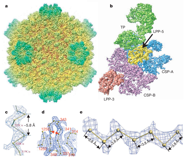
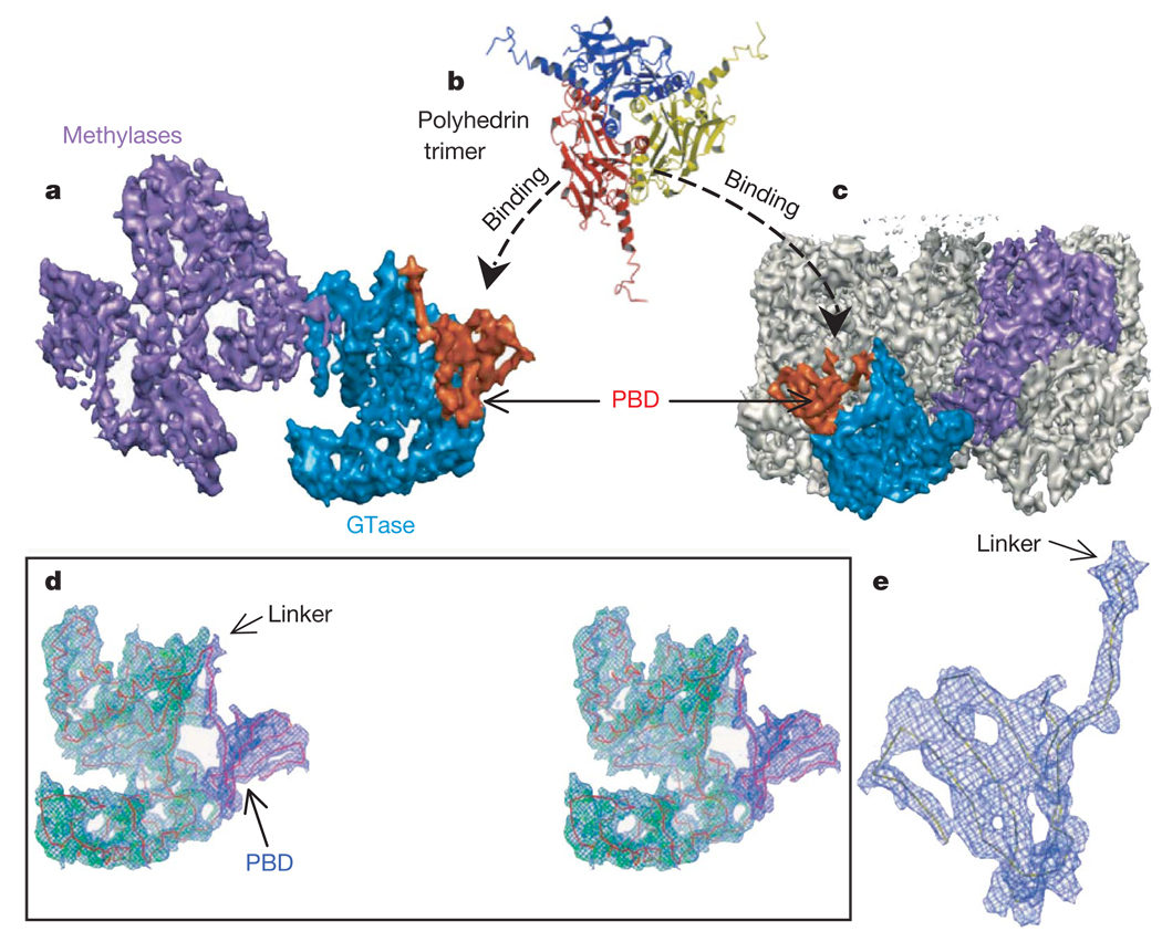
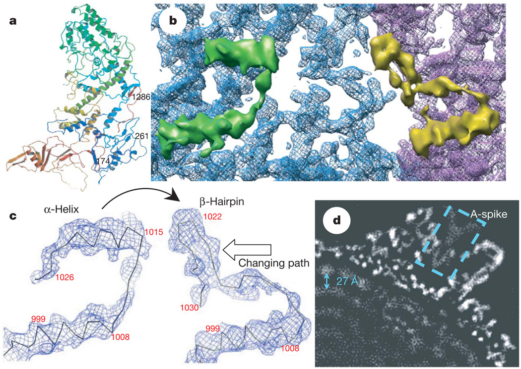
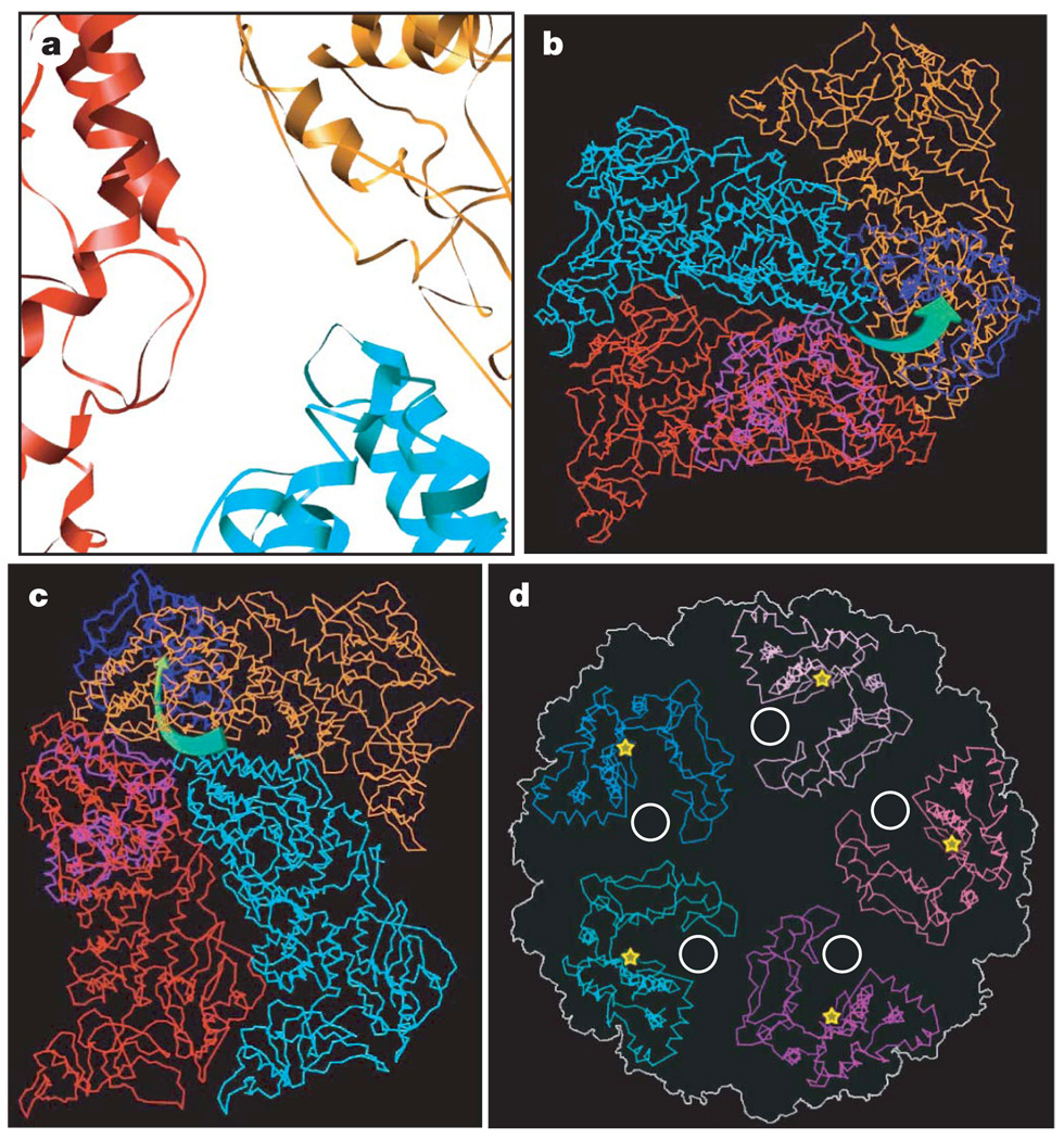
Cytoplasmic polyhedrosis virus (CPV) is unique within the Reoviridae family in having a turreted single-layer capsid contained within polyhedrin inclusion bodies, yet being fully capable of cell entry and endogenous RNA transcription. Biochemical data have shown that the amino-terminal 79 residues of the CPV turret protein (TP) is sufficient to bring CPV or engineered proteins into the polyhedrin matrix for micro-encapsulation. Here we report the three-dimensional structure of CPV at 3.88 A resolution using single-particle cryo-electron microscopy. Our map clearly shows the turns and deep grooves of alpha-helices, the strand separation in beta-sheets, and densities for loops and many bulky side chains; thus permitting atomic model-building effort from cryo-electron microscopy maps. We observed a helix-to-beta-hairpin conformational change between the two conformational states of the capsid shell protein in the region directly interacting with genomic RNA. We have also discovered a messenger RNA release hole coupled with the mRNA capping machinery unique to CPV. Furthermore, we have identified the polyhedrin-binding domain, a structure that has potential in nanobiotechnology applications.
细胞质多角体病毒(CPV)在呼肠孤病毒科中独具特色,其具有包含在多角体蛋白包涵体内的带炮塔的单层衣壳,但仍完全具备进入细胞和进行内源性RNA转录的能力。生化数据表明,CPV炮塔蛋白(TP)的氨基末端79个残基足以将CPV或工程蛋白带入多角体蛋白基质中进行微囊化。在此,我们使用单颗粒冷冻电子显微镜以3.88埃的分辨率报告了CPV的三维结构。我们的图谱清晰地显示了α螺旋的转角和深沟、β折叠中的链分离以及环和许多大侧链的密度;从而允许从冷冻电子显微镜图谱构建原子模型。我们在衣壳壳蛋白与基因组RNA直接相互作用的区域中观察到两种构象状态之间的螺旋到β发夹的构象变化。我们还发现了一个与CPV特有的mRNA加帽机制相关的信使RNA释放孔。此外,我们确定了多角体蛋白结合结构域,该结构在纳米生物技术应用中具有潜力。