• 文献检索
  • 文档翻译
  • 深度研究
  • 学术资讯
  • Suppr Zotero 插件Zotero 插件
  • 邀请有礼
  • 套餐&价格
  • 历史记录
应用&插件
Suppr Zotero 插件Zotero 插件浏览器插件Mac 客户端Windows 客户端微信小程序
定价
高级版会员购买积分包购买API积分包
服务
文献检索文档翻译深度研究API 文档MCP 服务
关于我们
关于 Suppr公司介绍联系我们用户协议隐私条款
关注我们

Suppr 超能文献

核心技术专利:CN118964589B侵权必究
粤ICP备2023148730 号-1Suppr @ 2026

文献检索

告别复杂PubMed语法,用中文像聊天一样搜索,搜遍4000万医学文献。AI智能推荐,让科研检索更轻松。

立即免费搜索

文件翻译

保留排版,准确专业,支持PDF/Word/PPT等文件格式,支持 12+语言互译。

免费翻译文档

深度研究

AI帮你快速写综述,25分钟生成高质量综述,智能提取关键信息,辅助科研写作。

立即免费体验

目的:颅骨形态的三维分析。

Objective three-dimensional analysis of cranial morphology.

作者信息

Marcus Jeffrey R, Domeshek Leahthan F, Das Rajesh, Marshall Sean, Nightingale Roger, Stokes Tracey H, Mukundan Srinivasan

机构信息

Interdisciplinary Craniofacial Imaging Lab, Duke University, Durham, NC, USA.

出版信息

Eplasty. 2008 Apr 9;8:e20.

PMID:18464892
原文链接:https://pmc.ncbi.nlm.nih.gov/articles/PMC2323201/
Abstract

OBJECTIVE

The lack of adequate means to objectively characterize cranial shape contributes to ongoing controversies in the surgical management of craniosynostosis. Cranial shape analysis must address relevant clinical questions objectively and thoroughly and must be broadly applicable across the spectrum of normal and abnormal. Herein, we demonstrate and statistically validate an automated computed tomography (CT)-based application for 3-dimensional characterization of skull morphology. The technology is intended for application to diagnostic imaging, surgical planning, and outcomes assessment.

METHODS

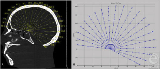
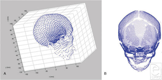
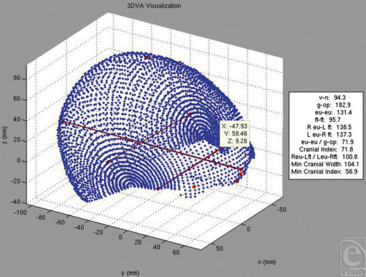
Three-dimensional vector analysis (3DVA) was applied to craniofacial CT data, generating three-dimensional cranial surface point clouds.

VALIDATION

To assess accuracy, measurements derived from the 3DVA analysis of a CT scan of a skull phantom were compared to those made directly from the Digital Imaging and Communications in Medicine data on a Vitrea workstation. To assess reproducibility, 3 readers independently analyzed human head CT scans using 3DVA.

APPLICATION

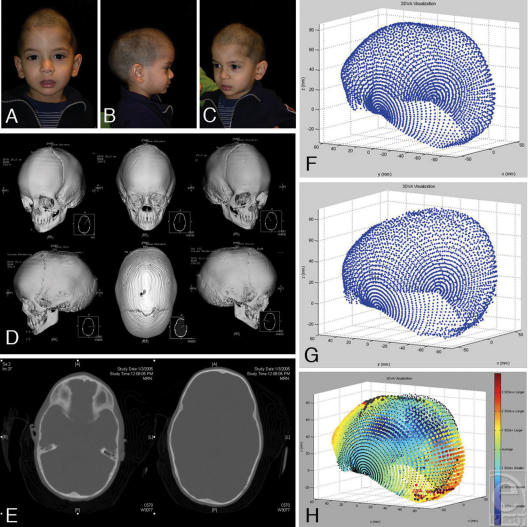
A normative database of 86 age-incremental pediatric patients was created. Preoperative craniosynostosis case datasets were analyzed using 3DVA and were compared with age-matched normative datasets.

RESULTS

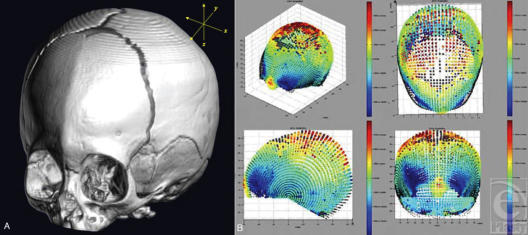
Accuracy and reproducibility of less than 1% mean error and less than 0.5 mm standard error in all cases validated 3DVA-derived distances. Three-dimensional vector analysis point clouds provide qualitative and quantitative representations of morphology. Regional dysmorphology in craniosynostosis cases is demonstrated graphically.

CONCLUSIONS

Three-dimensional vector analysis generated accurate, reproducible, and comprehensive craniofacial morphometric data. 3DVA may be used for paired data analysis (eg, a single subject undergoing surgical correction), comparative group data analysis, and craniofacial data archiving. The technique can provide objective characterization of craniofacial morphology previously not possible.

摘要

目的

缺乏客观描述颅骨形状的适当方法,这导致颅缝早闭手术治疗中持续存在争议。颅骨形状分析必须客观、全面地解决相关临床问题,并且必须广泛适用于正常和异常范围。在此,我们展示并通过统计学验证了一种基于计算机断层扫描(CT)的自动化应用程序,用于颅骨形态的三维表征。该技术旨在应用于诊断成像、手术规划和结果评估。

方法

将三维矢量分析(3DVA)应用于颅面CT数据,生成三维颅骨表面点云。

验证

为评估准确性,将从颅骨模型CT扫描的3DVA分析得出的测量结果与在Vitrea工作站上直接从医学数字成像和通信数据得出的测量结果进行比较。为评估可重复性,3名读者使用3DVA独立分析人头CT扫描。

应用

创建了一个包含86例年龄递增的儿科患者的规范数据库。使用3DVA分析术前颅缝早闭病例数据集,并与年龄匹配的规范数据集进行比较。

结果

在所有病例中,平均误差小于1%且标准误差小于0.5毫米的准确性和可重复性验证了3DVA得出的距离。三维矢量分析点云提供了形态的定性和定量表示。颅缝早闭病例中的区域畸形通过图形展示。

结论

三维矢量分析生成了准确、可重复且全面的颅面形态测量数据。3DVA可用于配对数据分析(例如,接受手术矫正的单个受试者)、比较组数据分析和颅面数据存档。该技术可以提供以前无法实现的颅面形态的客观表征。

https://cdn.ncbi.nlm.nih.gov/pmc/blobs/12b8/2323201/3b15d60fd106/eplasty08e20_fig6.jpg
https://cdn.ncbi.nlm.nih.gov/pmc/blobs/12b8/2323201/f733fd521b61/eplasty08e20_fig1.jpg
https://cdn.ncbi.nlm.nih.gov/pmc/blobs/12b8/2323201/3030876fb887/eplasty08e20_fig2.jpg
https://cdn.ncbi.nlm.nih.gov/pmc/blobs/12b8/2323201/b577b5865353/eplasty08e20_fig3.jpg
https://cdn.ncbi.nlm.nih.gov/pmc/blobs/12b8/2323201/0c2cfa6c8f00/eplasty08e20_fig4.jpg
https://cdn.ncbi.nlm.nih.gov/pmc/blobs/12b8/2323201/5002f078322d/eplasty08e20_fig5.jpg
https://cdn.ncbi.nlm.nih.gov/pmc/blobs/12b8/2323201/3b15d60fd106/eplasty08e20_fig6.jpg

相似文献

1
Objective three-dimensional analysis of cranial morphology.目的:颅骨形态的三维分析。
Eplasty. 2008 Apr 9;8:e20.
2
Use of a three-dimensional, normative database of pediatric craniofacial morphology for modern anthropometric analysis.利用儿童颅面形态的三维规范化数据库进行现代人体测量分析。
Plast Reconstr Surg. 2009 Dec;124(6):2076-2084. doi: 10.1097/PRS.0b013e3181bf7e1b.
3
Quantitative and qualitative assessment of morphology in sagittal synostosis: mid-sagittal vector analysis.矢状缝早闭形态学的定量与定性评估:矢状面矢量分析
J Craniofac Surg. 2006 Jul;17(4):680-6. doi: 10.1097/00001665-200607000-00013.
4
The metopic index: an anthropometric index for the quantitative assessment of trigonocephaly from metopic synostosis.额缝指数:一种用于定量评估额缝早闭所致三角头畸形的人体测量学指标。
J Neurosurg Pediatr. 2016 Sep;18(3):275-80. doi: 10.3171/2016.2.PEDS15524. Epub 2016 May 6.
5
Radiation-free quantification of head malformations in craniosynostosis patients from 3D photography.通过三维摄影对颅缝早闭患者头部畸形进行无辐射量化分析。
Proc SPIE Int Soc Opt Eng. 2018 Feb;10575. doi: 10.1117/12.2295374. Epub 2018 Feb 27.
6
Quantification of Head Shape from Three-Dimensional Photography for Presurgical and Postsurgical Evaluation of Craniosynostosis.三维摄影对头形的定量分析在颅缝早闭的术前和术后评估中的应用。
Plast Reconstr Surg. 2019 Dec;144(6):1051e-1060e. doi: 10.1097/PRS.0000000000006260.
7
Using Black Bone Magnetic Resonance Imaging in Craniofacial Virtual Surgical Planning: A Comparative Cadaver Study.颅颌面虚拟手术规划中使用黑骨磁共振成像:一项比较尸体研究。
Plast Reconstr Surg. 2018 Jun;141(6):1459-1470. doi: 10.1097/PRS.0000000000004396.
8
Objectification of cranial vault correction for craniosynostosis by three-dimensional photography.通过三维摄影实现颅盖畸形矫正的客观化。
J Craniomaxillofac Surg. 2012 Dec;40(8):726-30. doi: 10.1016/j.jcms.2012.01.007. Epub 2012 Feb 23.
9
The Natural History of Cranial Morphology in Sagittal Craniosynostosis.矢状缝早闭症中颅骨形态的自然史。
J Craniofac Surg. 2018 Jun;29(4):852-855. doi: 10.1097/SCS.0000000000004298.
10
Creating a normative database of age-specific 3D geometrical data, bone density, and bone thickness of the developing skull: a pilot study.建立发育中颅骨特定年龄的三维几何数据、骨密度和骨厚度的标准数据库:一项初步研究。
J Neurosurg Pediatr. 2015 Dec;16(6):687-702. doi: 10.3171/2015.4.PEDS1493. Epub 2015 Sep 4.

引用本文的文献

1
Normscan: open-source Python software to create average models from CT scans.Normscan:用于从CT扫描创建平均模型的开源Python软件。
Int J Comput Assist Radiol Surg. 2025 Jan;20(1):157-165. doi: 10.1007/s11548-024-03185-0. Epub 2024 May 20.
2
The efficacy of computed tomography scanning versus surface scanning in 3D finite element analysis.计算机断层扫描与表面扫描在三维有限元分析中的效果比较。
PeerJ. 2022 Aug 25;10:e13760. doi: 10.7717/peerj.13760. eCollection 2022.
3
Three-dimensional surface scanning methods in osteology: A topographical and geometric morphometric comparison.

本文引用的文献

1
Identifying reproducible patterns of calvarial dysmorphology in nonsyndromic sagittal craniosynostosis may affect operative intervention and outcomes assessment.识别非综合征性矢状缝早闭中可重复的颅骨畸形模式可能会影响手术干预和结果评估。
Plast Reconstr Surg. 2007 Apr 15;119(5):1546-1552. doi: 10.1097/01.prs.0000256067.42651.30.
2
Roentgenological skull measurements and their diagnostic applications.X线颅骨测量及其诊断应用。
Am J Roentgenol Radium Ther Nucl Med. 1952 Feb;67(2):197-209.
3
Spring-mediated cranioplasty compared with the modified pi-plasty for sagittal synostosis.
骨骼学中的三维表面扫描方法:地形学和几何形态测量学的比较。
Am J Phys Anthropol. 2021 Apr;174(4):846-858. doi: 10.1002/ajpa.24204. Epub 2021 Jan 7.
4
Cranial shape comparison for automated objective 3D craniosynostosis surgery planning.颅形比较用于自动化的客观 3D 颅缝早闭手术规划。
Sci Rep. 2018 Feb 20;8(1):3349. doi: 10.1038/s41598-018-21662-w.
5
Head circumference - a useful single parameter for skull volume development in cranial growth analysis?头围——颅骨生长分析中评估颅骨容积发育的有用单一参数?
Head Face Med. 2018 Jan 10;14(1):3. doi: 10.1186/s13005-017-0159-8.
6
Evaluation of morphological changes in the adult skull with age and sex.评估成年颅骨随年龄和性别的形态变化。
J Anat. 2016 Dec;229(6):838-846. doi: 10.1111/joa.12247. Epub 2014 Nov 18.
7
Cephalic index in the first three years of life: study of children with normal brain development based on computed tomography.生命前三年的头指数:基于计算机断层扫描对脑发育正常儿童的研究
ScientificWorldJournal. 2014 Feb 4;2014:502836. doi: 10.1155/2014/502836. eCollection 2014.
8
Validation of cephalic index measurements in scaphocephaly.舟状头畸形中头指数测量的验证
Childs Nerv Syst. 2013 Jun;29(6):1007-14. doi: 10.1007/s00381-013-2059-y. Epub 2013 Mar 7.
9
3D digital stereophotogrammetry: a practical guide to facial image acquisition.3D 数字体视摄影测量技术:面部图像采集实用指南。
Head Face Med. 2010 Jul 28;6:18. doi: 10.1186/1746-160X-6-18.
弹簧介导颅骨成形术与改良“π”形手术治疗矢状缝早闭的比较
Scand J Plast Reconstr Surg Hand Surg. 2003;37(4):208-15. doi: 10.1080/02844310310001823.
4
The reversal exchange technique of total calvarial reconstruction for sagittal synostosis.矢状缝早闭全颅骨重建的翻转交换技术
Plast Reconstr Surg. 2001 Jun;107(7):1637-46. doi: 10.1097/00006534-200106000-00001.
5
Clinical outcome of the modified pi-plasty procedure for sagittal synostosis.矢状缝早闭改良Pi成形术的临床疗效
J Craniofac Surg. 2001 May;12(3):218-24; discussion 225-6. doi: 10.1097/00001665-200105000-00003.
6
Sagittal synostosis: results of surgical treatment in 210 patients.矢状缝早闭:210例患者的手术治疗结果
Childs Nerv Syst. 2001 Jan;17(1-2):64-8. doi: 10.1007/s003810000353.
7
Sagittal craniosynostosis outcome assessment for two methods and timings of intervention.两种干预方法和时机的矢状缝早闭结局评估
Plast Reconstr Surg. 1999 May;103(6):1574-84. doi: 10.1097/00006534-199905060-00004.
8
Surgical management of sagittal synostosis: a comparative analysis of strip craniectomy and calvarial vault remodeling.矢状缝早闭的手术治疗:条状颅骨切除术与颅骨穹窿重塑的对比分析
Pediatr Neurosurg. 1997 Sep;27(3):137-48. doi: 10.1159/000121241.
9
Sagittal synostosis: quantitative assessment of presenting deformity and surgical results based on CT scans.矢状缝早闭:基于CT扫描对呈现的畸形和手术结果进行定量评估。
Plast Reconstr Surg. 1993 Nov;92(6):1015-24; discussion 1225-6.
10
Correction of scaphocephaly secondary to ventricular shunting procedures.脑室分流术后舟状头畸形的矫正
Plast Reconstr Surg. 1995 Oct;96(5):1012-9. doi: 10.1097/00006534-199510000-00002.