• 文献检索
  • 文档翻译
  • 深度研究
  • 学术资讯
  • Suppr Zotero 插件Zotero 插件
  • 邀请有礼
  • 套餐&价格
  • 历史记录
应用&插件
Suppr Zotero 插件Zotero 插件浏览器插件Mac 客户端Windows 客户端微信小程序
定价
高级版会员购买积分包购买API积分包
服务
文献检索文档翻译深度研究API 文档MCP 服务
关于我们
关于 Suppr公司介绍联系我们用户协议隐私条款
关注我们

Suppr 超能文献

核心技术专利:CN118964589B侵权必究
粤ICP备2023148730 号-1Suppr @ 2026

文献检索

告别复杂PubMed语法,用中文像聊天一样搜索,搜遍4000万医学文献。AI智能推荐,让科研检索更轻松。

立即免费搜索

文件翻译

保留排版,准确专业,支持PDF/Word/PPT等文件格式,支持 12+语言互译。

免费翻译文档

深度研究

AI帮你快速写综述,25分钟生成高质量综述,智能提取关键信息,辅助科研写作。

立即免费体验

C57BL/10小鼠和mdx小鼠间充质干细胞的比较研究。

Comparative study of mesenchymal stem cells from C57BL/10 and mdx mice.

作者信息

Li Yong, Zhang Cheng, Xiong Fu, Yu Mei-juan, Peng Fu-Lin, Shang Yan-chang, Zhao Cui-ping, Xu Yong-feng, Liu Zheng-shan, Zhou Chang, Wu Jin-lang

机构信息

Department of Neurology, the First Affiliated Hospital, Sun Yat-sen University, Guangzhou, Guangdong, ProC.

出版信息

BMC Cell Biol. 2008 May 19;9:24. doi: 10.1186/1471-2121-9-24.

DOI:10.1186/1471-2121-9-24
PMID:18489762
原文链接:https://pmc.ncbi.nlm.nih.gov/articles/PMC2415111/
Abstract

BACKGROUND

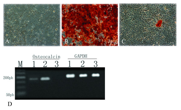
Human mesenchymal stem cells (MSCs) have been studied and applied extensively because of their ability to self-renew and differentiate into various cell types. Since most human diseases models are murine, mouse MSCs should have been studied in detail. The mdx mouse - a Duchenne muscular dystrophy model - was produced by introducing a point mutation in the dystrophin gene. To understand the role of dystrophin in MSCs, we compared MSCs from mdx and C57BL/10 mice, focusing particularly on the aspects of light and electron microscopic morphology, immunophenotyping, and differentiation potential.

RESULTS

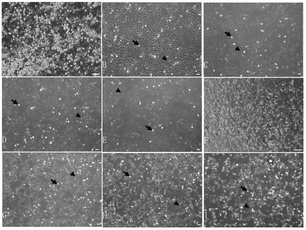
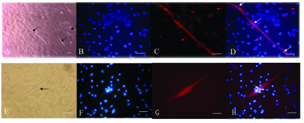
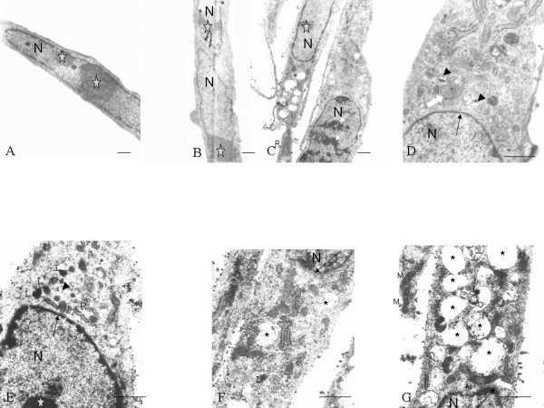
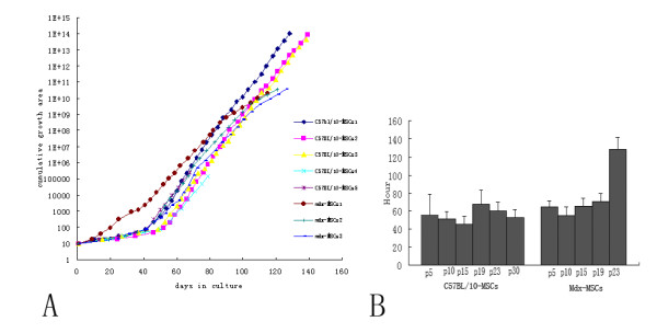
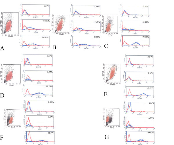
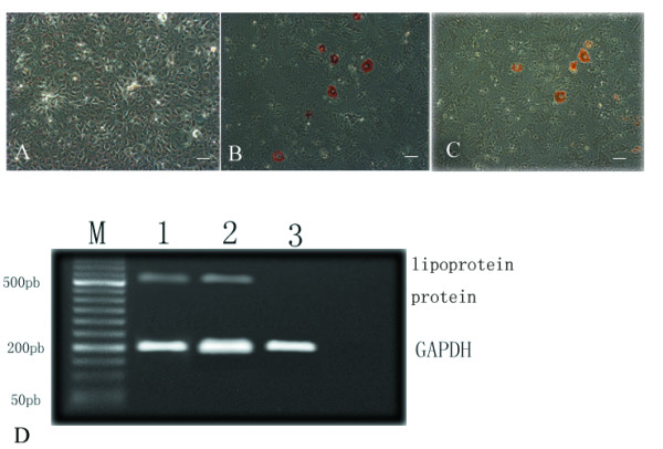
Our study showed that at passage 10, mdx-MSCs exhibited increased heterochromatin, larger vacuoles, and more lysosomes under electron microscopy compared to C57BL/10-MSCs. C57BL/10-MSCs formed a few myotubes, while mdx-MSCs did not at the same passages. By passage 21, mdx-MSCs but not C57BL/10-MSCs had gradually lost their proliferative ability. In addition, a significant difference in the expression of CD34, not Sca-1 and CD11b, was observed between the MSCs from the 2 mice.

CONCLUSION

Our current study reveals that the MSCs from the 2 mice, namely, C57BL/10 and mdx, exhibit differences in proliferative and myogenic abilities. The results suggest that the changes in mouse MSC behavior may be influenced by lack of dystrophin protein in mdx mouse.

摘要

背景

人间充质干细胞(MSCs)因其自我更新和分化为多种细胞类型的能力而得到广泛研究和应用。由于大多数人类疾病模型是鼠类的,因此应该对小鼠间充质干细胞进行详细研究。mdx小鼠——一种杜氏肌营养不良模型——是通过在肌营养不良蛋白基因中引入点突变产生的。为了了解肌营养不良蛋白在间充质干细胞中的作用,我们比较了mdx小鼠和C57BL/10小鼠的间充质干细胞,特别关注光镜和电镜形态学、免疫表型分析以及分化潜能等方面。

结果

我们的研究表明,在第10代时,与C57BL/10间充质干细胞相比,mdx间充质干细胞在电镜下表现出异染色质增加、空泡更大和溶酶体更多。C57BL/10间充质干细胞形成了一些肌管,而mdx间充质干细胞在相同代次时未形成。到第21代时,mdx间充质干细胞逐渐失去了增殖能力,而C57BL/10间充质干细胞没有。此外,在这两种小鼠的间充质干细胞之间观察到CD34表达存在显著差异,而Sca-1和CD11b表达无差异。

结论

我们目前的研究表明,C57BL/10和mdx这两种小鼠的间充质干细胞在增殖和生肌能力方面存在差异。结果表明,mdx小鼠中肌营养不良蛋白的缺乏可能影响小鼠间充质干细胞的行为变化。

https://cdn.ncbi.nlm.nih.gov/pmc/blobs/8bb1/2415111/954f047b9f7e/1471-2121-9-24-7.jpg
https://cdn.ncbi.nlm.nih.gov/pmc/blobs/8bb1/2415111/92a3944e7b45/1471-2121-9-24-1.jpg
https://cdn.ncbi.nlm.nih.gov/pmc/blobs/8bb1/2415111/18d8b8b24c1e/1471-2121-9-24-2.jpg
https://cdn.ncbi.nlm.nih.gov/pmc/blobs/8bb1/2415111/99aa979de02d/1471-2121-9-24-5.jpg
https://cdn.ncbi.nlm.nih.gov/pmc/blobs/8bb1/2415111/ae3b7baea731/1471-2121-9-24-3.jpg
https://cdn.ncbi.nlm.nih.gov/pmc/blobs/8bb1/2415111/29327f84d077/1471-2121-9-24-4.jpg
https://cdn.ncbi.nlm.nih.gov/pmc/blobs/8bb1/2415111/7bdb0ffbdd81/1471-2121-9-24-6.jpg
https://cdn.ncbi.nlm.nih.gov/pmc/blobs/8bb1/2415111/954f047b9f7e/1471-2121-9-24-7.jpg

相似文献

1
Comparative study of mesenchymal stem cells from C57BL/10 and mdx mice.C57BL/10小鼠和mdx小鼠间充质干细胞的比较研究。
BMC Cell Biol. 2008 May 19;9:24. doi: 10.1186/1471-2121-9-24.
2
Transplantation of Dystrophin Expressing Chimeric Human Cells of Myoblast/Mesenchymal Stem Cell Origin Improves Function in Duchenne Muscular Dystrophy Model.肌母细胞/间充质干细胞来源的表达抗肌萎缩蛋白嵌合人细胞移植可改善杜氏肌营养不良症模型的功能。
Stem Cells Dev. 2021 Feb;30(4):190-202. doi: 10.1089/scd.2020.0161. Epub 2021 Jan 22.
3
Cardiac Protection after Systemic Transplant of Dystrophin Expressing Chimeric (DEC) Cells to the mdx Mouse Model of Duchenne Muscular Dystrophy.系统性移植表达抗肌萎缩蛋白嵌合(DEC)细胞后对 mdx 小鼠模型杜氏肌营养不良症的心脏保护作用。
Stem Cell Rev Rep. 2019 Dec;15(6):827-841. doi: 10.1007/s12015-019-09916-0.
4
A comparative study of bone marrow mesenchymal stem cell functionality in C57BL and mdx mice.C57BL 和 mdx 小鼠骨髓间充质干细胞功能的比较研究。
Neurosci Lett. 2012 Aug 15;523(2):139-44. doi: 10.1016/j.neulet.2012.06.061. Epub 2012 Jul 3.
5
[Mice adipose derived Flk-1+ mesenchymal stem cells can ameliorate Duchenne's muscular dystrophy in Mdx mice for their multilineage potential].[小鼠脂肪来源的Flk-1+间充质干细胞因其多向分化潜能可改善Mdx小鼠的杜氏肌营养不良症]
Zhongguo Shi Yan Xue Ye Xue Za Zhi. 2007 Apr;15(2):306-12.
6
[Construction of recombinant adenovirus including microdystrophin and expression in the mesenchymal cells of mdx mice].[包含微肌营养不良蛋白的重组腺病毒构建及其在mdx小鼠间充质细胞中的表达]
Sheng Wu Gong Cheng Xue Bao. 2007 Jan;23(1):27-32. doi: 10.1016/s1872-2075(07)60003-x.
7
Impaired regenerative capacity and lower revertant fibre expansion in dystrophin-deficient mdx muscles on DBA/2 background.在 DBA/2 背景下,缺乏 dystrophin 的 mdx 肌肉的再生能力受损,回复纤维扩张减少。
Sci Rep. 2016 Dec 7;6:38371. doi: 10.1038/srep38371.
8
Multipotential mesoangioblast stem cell therapy in the mdx/utrn-/- mouse model for Duchenne muscular dystrophy.多能性中血管周细胞干细胞疗法在杜氏肌营养不良症的mdx/utrn-/-小鼠模型中的应用
Regen Med. 2007 May;2(3):275-88. doi: 10.2217/17460751.2.3.275.
9
[Mesenchymal stem cells transplanted in mdx mice differentiate into myocytes and express dystrophin/utrophin].移植到mdx小鼠体内的间充质干细胞分化为肌细胞并表达抗肌萎缩蛋白/抗肌萎缩蛋白相关蛋白
Nan Fang Yi Ke Da Xue Xue Bao. 2009 May;29(5):974-8.
10
Mesenchymal stem cells derived from human induced pluripotent stem cells improve the engraftment of myogenic cells by secreting urokinase-type plasminogen activator receptor (uPAR).人诱导多能干细胞来源的间充质干细胞通过分泌尿激酶型纤溶酶原激活物受体 (uPAR) 改善成肌细胞的植入。
Stem Cell Res Ther. 2021 Oct 9;12(1):532. doi: 10.1186/s13287-021-02594-1.

引用本文的文献

1
Manufacturing exosomes for wound healing: Comparative analysis of culture media.用于伤口愈合的外泌体的制造:培养基的比较分析。
PLoS One. 2024 Nov 14;19(11):e0313697. doi: 10.1371/journal.pone.0313697. eCollection 2024.
2
Labeling and Tracking of Individual Human Mesenchymal Stromal Cells Using Photoconvertible Fluorescent Microcapsules.利用光转化荧光微胶囊对个体人类间充质基质细胞进行标记和追踪。
Int J Mol Sci. 2023 Sep 4;24(17):13665. doi: 10.3390/ijms241713665.
3
Ultrastructure Morphological Characterization of Different Passages of Rat Dental Follicle Stem Cells at Culture.

本文引用的文献

1
CD34 expression on murine marrow-derived mesenchymal stromal cells: impact on neovascularization.小鼠骨髓来源间充质基质细胞上的CD34表达:对新血管形成的影响
Exp Hematol. 2008 Jan;36(1):93-103. doi: 10.1016/j.exphem.2007.08.032. Epub 2007 Nov 26.
2
Human multipotent adipose-derived stem cells restore dystrophin expression of Duchenne skeletal-muscle cells in vitro.人多能脂肪来源干细胞在体外恢复杜氏骨骼肌细胞的肌营养不良蛋白表达。
Biol Cell. 2008 Apr;100(4):231-41. doi: 10.1042/BC20070102.
3
Menstrual blood-derived cells confer human dystrophin expression in the murine model of Duchenne muscular dystrophy via cell fusion and myogenic transdifferentiation.
大鼠牙囊干细胞不同传代培养时的超微结构形态学特征
J Microsc Ultrastruct. 2019 Apr-Jun;7(2):57-64. doi: 10.4103/JMAU.JMAU_44_18.
4
DP71 and SERCA2 alteration in human neurons of a Duchenne muscular dystrophy patient.杜氏肌营养不良症患者的人类神经元中 DP71 和 SERCA2 的改变。
Stem Cell Res Ther. 2019 Jan 15;10(1):29. doi: 10.1186/s13287-018-1125-5.
5
Mesenchymal Stem Cells of Different Origin-Seeded Bioceramic Construct in Regeneration of Bone Defect in Rabbit.不同来源间充质干细胞接种生物陶瓷支架修复兔骨缺损的研究
Tissue Eng Regen Med. 2018 Jul 10;15(4):477-492. doi: 10.1007/s13770-018-0129-7. eCollection 2018 Aug.
6
Transgene expression and differentiation of baculovirus-transduced adipose-derived stem cells from dystrophin-utrophin double knock-out mouse.来自肌营养不良蛋白-促肌萎缩蛋白双敲除小鼠的杆状病毒转导脂肪来源干细胞的转基因表达与分化
Neural Regen Res. 2012 Aug 5;7(22):1695-702. doi: 10.3969/j.issn.1673-5374.2012.22.003.
7
Mesenchymal stem cells as cellular vectors for pediatric neurological disorders.间充质干细胞作为小儿神经疾病的细胞载体
Brain Res. 2014 Jul 21;1573:92-107. doi: 10.1016/j.brainres.2014.05.029. Epub 2014 May 22.
8
Evaluation of neural damage in Duchenne muscular dystrophy patients.杜氏肌营养不良症患者神经损伤的评估。
Acta Myol. 2014 May;33(1):13-8.
9
Enhancing the efficacy of mesenchymal stem cell therapy.提高间充质干细胞疗法的疗效。
World J Stem Cells. 2014 Apr 26;6(2):82-93. doi: 10.4252/wjsc.v6.i2.82.
10
Isolation, characterization and the multi-lineage differentiation potential of rabbit bone marrow-derived mesenchymal stem cells.兔骨髓间充质干细胞的分离、鉴定及其多向分化潜能。
J Anat. 2013 Apr;222(4):437-50. doi: 10.1111/joa.12032.
月经血源性细胞通过细胞融合和肌源性转分化在杜兴氏肌营养不良症小鼠模型中赋予人抗肌萎缩蛋白表达。
Mol Biol Cell. 2007 May;18(5):1586-94. doi: 10.1091/mbc.e06-09-0872. Epub 2007 Feb 21.
4
Widespread distribution and muscle differentiation of human fetal mesenchymal stem cells after intrauterine transplantation in dystrophic mdx mouse.
Stem Cells. 2007 Apr;25(4):875-84. doi: 10.1634/stemcells.2006-0694. Epub 2006 Dec 21.
5
Stem cells from umbilical cord blood differentiate into myotubes and express dystrophin in vitro only after exposure to in vivo muscle environment.来自脐带血的干细胞仅在暴露于体内肌肉环境后才会在体外分化为肌管并表达抗肌萎缩蛋白。
Biol Cell. 2007 Apr;99(4):185-96. doi: 10.1042/BC20060075.
6
Mesenchymal stromal cells.间充质基质细胞
Curr Opin Hematol. 2006 Nov;13(6):419-25. doi: 10.1097/01.moh.0000245697.54887.6f.
7
The mesenchymal stem cell advantage.间充质干细胞的优势。
Stem Cells Dev. 2006 Aug;15(4):473-4. doi: 10.1089/scd.2006.15.473.
8
Murine mesenchymal stem cells isolated by low density primary culture system.通过低密度原代培养系统分离的小鼠间充质干细胞。
Dev Growth Differ. 2006 Aug;48(6):361-70. doi: 10.1111/j.1440-169X.2006.00874.x.
9
Mesenchymal stem cells in immunoregulation.间充质干细胞在免疫调节中的作用
Immunol Cell Biol. 2006 Oct;84(5):413-21. doi: 10.1111/j.1440-1711.2006.01458.x. Epub 2006 Jul 20.
10
Mesenchymal stem cells reside in virtually all post-natal organs and tissues.间充质干细胞几乎存在于所有出生后的器官和组织中。
J Cell Sci. 2006 Jun 1;119(Pt 11):2204-13. doi: 10.1242/jcs.02932. Epub 2006 May 9.