Brodoefel H, Reimann A, Heuschmid M, Tsiflikas I, Kopp A F, Schroeder S, Claussen C D, Clouse M E, Burgstahler C
Department of Diagnostic Radiology, Eberhard-Karls-University, Tübingen, Germany.
Eur Radiol. 2008 Nov;18(11):2466-74. doi: 10.1007/s00330-008-1019-5. Epub 2008 May 20.
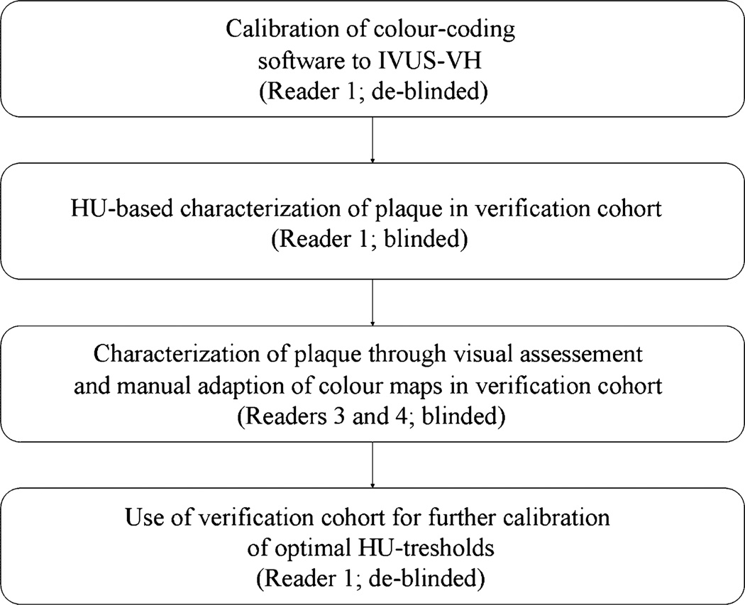
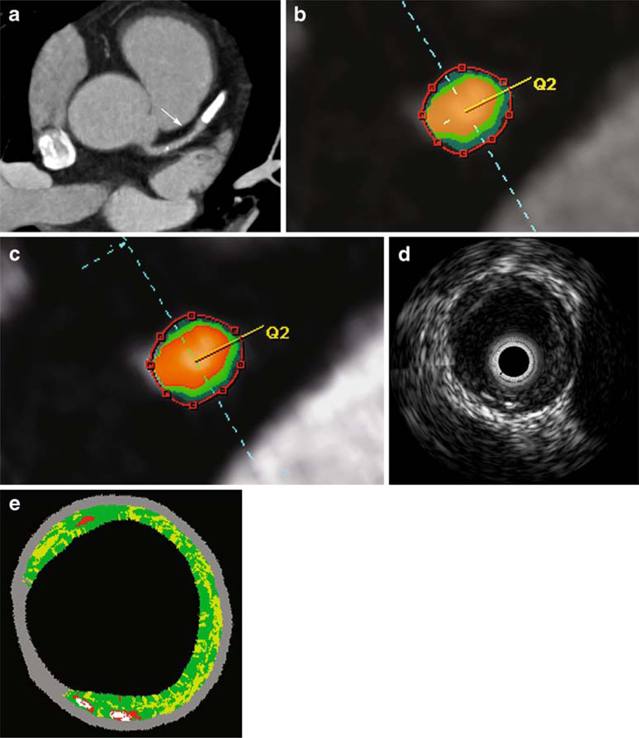
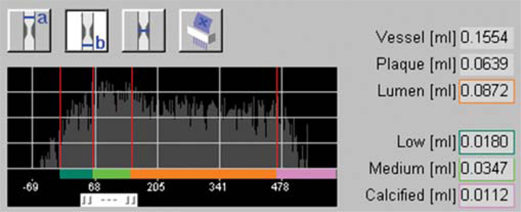
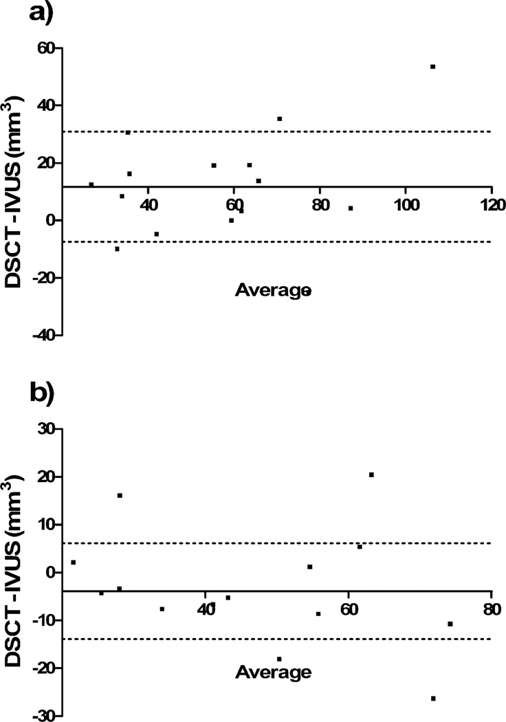
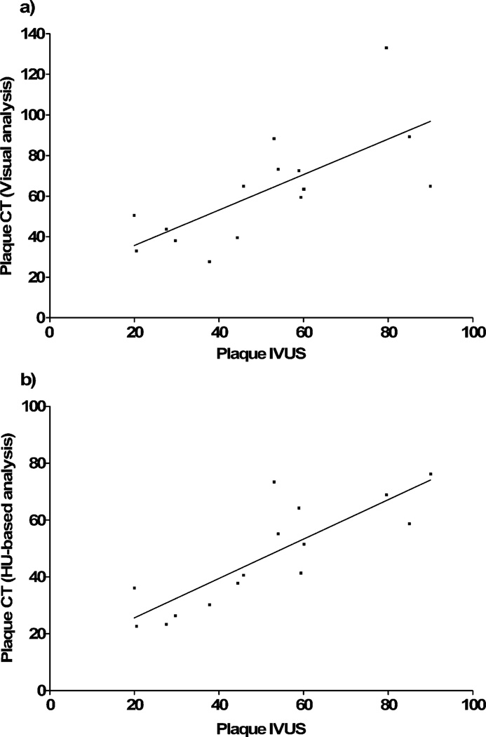
To assess HU-based color mapping for characterization of coronary plaque, using intravascular ultrasound virtual histology (IVUS-VH) as a standard of reference. Dual-source computed tomography and IVUS-VH were prospectively performed in 13 patients. In five lesions, HU thresholds of the color-coding software were calibrated to IVUS-VH. In a 15-lesion verification cohort, volumes of vessel, lumen and plaque or percentages of lipid, fibrous and calcified components were obtained through use of pre-set HU cut-offs as well as through purely visual adjustment of color maps. Calibrated HU ranges for fatty or fibrous plaque, lumen and calcification were -10-69, 70-158, 159-436 and 437+. Using these cut-offs, HU-based analysis achieved good agreement of plaque volume with IVUS (47.0 vs. 51.0 mm(3)). Visual segmentation led to significant overestimation of atheroma (61.6 vs. 51.0 mm(3); P = 0.04) Correlation coefficients for volumes of vessel, lumen and plaque were 0.92, 0.87 and 0.83 with HU-based analysis or 0.92, 0.85 and 0.71 with visual evaluation. With both methods, correlation of percentage plaque composition was poor or insignificant. HU-based plaque analysis showed good reproducibility with intra-class correlation coefficients being 0.90 for plaque volume and 0.81, 0.94 or 0.98 for percentages of fatty, fibrous or calcified components. With use of optimized HU thresholds, color mapping allows for accurate and reproducible quantification of coronary plaque.
以血管内超声虚拟组织学(IVUS-VH)作为参考标准,评估基于HU的彩色映射用于冠状动脉斑块特征分析的情况。对13例患者前瞻性地进行了双源计算机断层扫描和IVUS-VH检查。在5个病变中,将彩色编码软件的HU阈值校准为IVUS-VH。在一个15个病变的验证队列中,通过使用预设的HU截断值以及通过纯视觉调整彩色图谱,获得了血管、管腔和斑块的体积或脂质、纤维和钙化成分的百分比。脂肪或纤维斑块、管腔和钙化的校准HU范围分别为-10-69、70-158、159-436和437+。使用这些截断值,基于HU的分析与IVUS在斑块体积上取得了良好的一致性(47.0对51.0 mm³)。视觉分割导致对动脉粥样硬化的显著高估(61.6对51.0 mm³;P = 0.04)。基于HU的分析中,血管、管腔和斑块体积的相关系数分别为0.92、0.87和0.83,视觉评估的相关系数分别为0.92、0.85和0.71。两种方法中,斑块成分百分比的相关性都较差或不显著。基于HU的斑块分析显示出良好的可重复性,斑块体积的组内相关系数为0.90,脂肪、纤维或钙化成分百分比的组内相关系数分别为0.81、0.94或0.98。通过使用优化的HU阈值进行彩色映射,能够对冠状动脉斑块进行准确且可重复的定量分析。