• 文献检索
  • 文档翻译
  • 深度研究
  • 学术资讯
  • Suppr Zotero 插件Zotero 插件
  • 邀请有礼
  • 套餐&价格
  • 历史记录
应用&插件
Suppr Zotero 插件Zotero 插件浏览器插件Mac 客户端Windows 客户端微信小程序
定价
高级版会员购买积分包购买API积分包
服务
文献检索文档翻译深度研究API 文档MCP 服务
关于我们
关于 Suppr公司介绍联系我们用户协议隐私条款
关注我们

Suppr 超能文献

核心技术专利:CN118964589B侵权必究
粤ICP备2023148730 号-1Suppr @ 2026

文献检索

告别复杂PubMed语法,用中文像聊天一样搜索,搜遍4000万医学文献。AI智能推荐,让科研检索更轻松。

立即免费搜索

文件翻译

保留排版,准确专业,支持PDF/Word/PPT等文件格式,支持 12+语言互译。

免费翻译文档

深度研究

AI帮你快速写综述,25分钟生成高质量综述,智能提取关键信息,辅助科研写作。

立即免费体验

光学相干断层扫描的扫描圈位置及视网膜神经纤维层平均测量值的变异性

Optical coherence tomography scan circle location and mean retinal nerve fiber layer measurement variability.

作者信息

Gabriele Michelle L, Ishikawa Hiroshi, Wollstein Gadi, Bilonick Richard A, Townsend Kelly A, Kagemann Larry, Wojtkowski Maciej, Srinivasan Vivek J, Fujimoto James G, Duker Jay S, Schuman Joel S

机构信息

UPMC Eye Center, Eye and Ear Institute, Ophthalmology and Visual Science Research Center, Department of Ophthalmology, University of Pittsburgh School of Medicine, Pittsburgh, Pennsylvania 15213, USA.

出版信息

Invest Ophthalmol Vis Sci. 2008 Jun;49(6):2315-21. doi: 10.1167/iovs.07-0873.

DOI:10.1167/iovs.07-0873
PMID:18515577
原文链接:https://pmc.ncbi.nlm.nih.gov/articles/PMC2728289/
Abstract

PURPOSE

To investigate the effect on optical coherence tomography (OCT) retinal nerve fiber layer (RNFL) thickness measurements of varying the standard 3.4-mm-diameter circle location.

METHODS

The optic nerve head (ONH) region of 17 eyes of 17 healthy subjects was imaged with high-speed, ultrahigh-resolution OCT (hsUHR-OCT; 501 x 180 axial scans covering a 6 x 6-mm area; scan time, 3.84 seconds) for a comprehensive sampling. This method allows for systematic simulation of the variable circle placement effect. RNFL thickness was measured on this three-dimensional dataset by using a custom-designed software program. RNFL thickness was resampled along a 3.4-mm-diameter circle centered on the ONH, then along 3.4-mm circles shifted horizontally (x-shift), vertically (y-shift) and diagonally up to +/-500 microm (at 100-microm intervals). Linear mixed-effects models were used to determine RNFL thickness as a function of the scan circle shift. A model for the distance between the two thickest measurements along the RNFL thickness circular profile (peak distance) was also calculated.

RESULTS

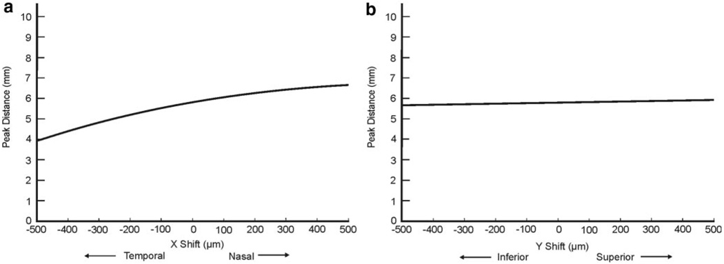
RNFL thickness tended to decrease with both positive and negative x- and y-shifts. The range of shifts that caused a decrease greater than the variability inherent to the commercial device was greater in both nasal and temporal quadrants than in the superior and inferior ones. The model for peak distance demonstrated that as the scan moves nasally, the RNFL peak distance increases, and as the circle moves temporally, the distance decreases. Vertical shifts had a minimal effect on peak distance.

CONCLUSIONS

The location of the OCT scan circle affects RNFL thickness measurements. Accurate registration of OCT scans is essential for measurement reproducibility and longitudinal examination (ClinicalTrials.gov number, NCT00286637).

摘要

目的

研究改变标准3.4毫米直径圆的位置对光学相干断层扫描(OCT)测量视网膜神经纤维层(RNFL)厚度的影响。

方法

对17名健康受试者的17只眼睛的视神经乳头(ONH)区域进行高速、超高分辨率OCT(hsUHR - OCT;501×180轴向扫描,覆盖6×6毫米区域;扫描时间3.84秒)成像,以进行全面采样。该方法允许系统模拟可变圆放置的效果。使用定制设计的软件程序在这个三维数据集中测量RNFL厚度。RNFL厚度首先沿以ONH为中心的3.4毫米直径圆进行重采样,然后沿水平(x轴偏移)、垂直(y轴偏移)和对角线方向最多偏移±500微米(以100微米间隔)的3.4毫米圆进行重采样。使用线性混合效应模型确定RNFL厚度作为扫描圆偏移的函数。还计算了沿RNFL厚度圆形轮廓的两个最厚测量值之间的距离模型(峰值距离)。

结果

RNFL厚度在x轴和y轴正负偏移时均趋于减小。导致下降幅度大于商用设备固有变异性的偏移范围在鼻侧和颞侧象限均大于上侧和下侧象限。峰值距离模型表明,随着扫描向鼻侧移动,RNFL峰值距离增加,随着圆向颞侧移动,距离减小。垂直偏移对峰值距离的影响最小。

结论

OCT扫描圆的位置会影响RNFL厚度测量。OCT扫描的准确配准对于测量的可重复性和纵向检查至关重要(ClinicalTrials.gov编号,NCT00286637)。

https://cdn.ncbi.nlm.nih.gov/pmc/blobs/08d0/2728289/bfb5c29ee30f/nihms84277f6.jpg
https://cdn.ncbi.nlm.nih.gov/pmc/blobs/08d0/2728289/0ed07f038992/nihms84277f1.jpg
https://cdn.ncbi.nlm.nih.gov/pmc/blobs/08d0/2728289/a645b48a04a6/nihms84277f2.jpg
https://cdn.ncbi.nlm.nih.gov/pmc/blobs/08d0/2728289/856cb6df5f93/nihms84277f3.jpg
https://cdn.ncbi.nlm.nih.gov/pmc/blobs/08d0/2728289/65293b95921d/nihms84277f4.jpg
https://cdn.ncbi.nlm.nih.gov/pmc/blobs/08d0/2728289/534b5871ce9d/nihms84277f5.jpg
https://cdn.ncbi.nlm.nih.gov/pmc/blobs/08d0/2728289/bfb5c29ee30f/nihms84277f6.jpg

相似文献

1
Optical coherence tomography scan circle location and mean retinal nerve fiber layer measurement variability.光学相干断层扫描的扫描圈位置及视网膜神经纤维层平均测量值的变异性
Invest Ophthalmol Vis Sci. 2008 Jun;49(6):2315-21. doi: 10.1167/iovs.07-0873.
2
Peripapillary nerve fiber layer thickness profile determined with high speed, ultrahigh resolution optical coherence tomography high-density scanning.采用高速、超高分辨率光学相干断层扫描高密度扫描测定的视乳头周围神经纤维层厚度轮廓。
Invest Ophthalmol Vis Sci. 2007 Jul;48(7):3154-60. doi: 10.1167/iovs.06-1416.
3
Effect of improper scan alignment on retinal nerve fiber layer thickness measurements using Stratus optical coherence tomograph.使用Stratus光学相干断层扫描仪时,扫描对准不当对视网膜神经纤维层厚度测量的影响。
J Glaucoma. 2008 Aug;17(5):341-9. doi: 10.1097/IJG.0b013e31815c3aeb.
4
Custom measurement of retinal nerve fiber layer thickness using STRATUS OCT in normal eyes.在正常眼睛中使用STRATUS OCT进行视网膜神经纤维层厚度的定制测量。
Eur J Ophthalmol. 2005 May-Jun;15(3):360-6. doi: 10.1177/112067210501500308.
5
Thickness and birefringence of healthy retinal nerve fiber layer tissue measured with polarization-sensitive optical coherence tomography.采用偏振敏感光学相干断层扫描技术测量健康视网膜神经纤维层组织的厚度和双折射。
Invest Ophthalmol Vis Sci. 2004 Aug;45(8):2606-12. doi: 10.1167/iovs.03-1160.
6
Adjusted peripapillary retinal nerve fiber layer thickness measurements based on the optic nerve head scan angle.基于视神经盘扫描角度的调整后的视盘周围视网膜神经纤维层厚度测量。
Invest Ophthalmol Vis Sci. 2010 Aug;51(8):4067-74. doi: 10.1167/iovs.09-4301. Epub 2010 Mar 17.
7
Correlation between retinal nerve fibre layer thickness and optic nerve head size: an optical coherence tomography study.视网膜神经纤维层厚度与视神经乳头大小之间的相关性:一项光学相干断层扫描研究。
Br J Ophthalmol. 2005 Apr;89(4):489-92. doi: 10.1136/bjo.2004.052498.
8
Retinal nerve fiber layer thickness measurement comparability between time domain optical coherence tomography (OCT) and spectral domain OCT.时域光学相干断层扫描(OCT)和频域 OCT 测量视网膜神经纤维层厚度的可比性。
Invest Ophthalmol Vis Sci. 2010 Feb;51(2):896-902. doi: 10.1167/iovs.09-4110. Epub 2009 Sep 8.
9
Retinal nerve fiber layer assessment: area versus thickness measurements from elliptical scans centered on the optic nerve.视网膜神经纤维层评估:以视神经为中心的椭圆形扫描的面积与厚度测量。
Invest Ophthalmol Vis Sci. 2011 Apr 16;52(5):2477-89. doi: 10.1167/iovs.10-6105. Print 2011 Apr.
10
The effect of scan diameter on retinal nerve fiber layer thickness measurement using stratus optic coherence tomography.扫描直径对使用Stratus光学相干断层扫描测量视网膜神经纤维层厚度的影响。
Arch Ophthalmol. 2007 Jul;125(7):901-5. doi: 10.1001/archopht.125.7.901.

引用本文的文献

1
Influence of OSCAR-IB Criteria on Test-Retest Reliability of Cirrus HD-OCT Retinal Thickness Measurements in People With Multiple Sclerosis.OSCAR-IB标准对多发性硬化症患者Cirrus HD-OCT视网膜厚度测量的重测信度的影响
Neurol Neuroimmunol Neuroinflamm. 2025 Nov;12(6):e200458. doi: 10.1212/NXI.0000000000200458. Epub 2025 Aug 29.
2
Normative Variability in Retinal Nerve Fiber Layer Thickness: Does It Matter Where the Peaks Are?视网膜神经纤维层厚度的正常变异性:峰值位置重要吗?
Transl Vis Sci Technol. 2025 May 1;14(5):13. doi: 10.1167/tvst.14.5.13.
3
Interocular comparison of peripapillary retinal nerve fiber layer thickness and vasculature in non-pathological myopia with anisometropia.

本文引用的文献

1
Ultrahigh-resolution, high-speed, Fourier domain optical coherence tomography and methods for dispersion compensation.超高分辨率、高速傅里叶域光学相干断层扫描及色散补偿方法。
Opt Express. 2004 May 31;12(11):2404-22. doi: 10.1364/opex.12.002404.
2
Sensitivity advantage of swept source and Fourier domain optical coherence tomography.扫频光源和傅里叶域光学相干断层扫描的灵敏度优势。
Opt Express. 2003 Sep 8;11(18):2183-9. doi: 10.1364/oe.11.002183.
3
Performance of fourier domain vs. time domain optical coherence tomography.傅里叶域与时间域光学相干断层扫描的性能比较
非病理性屈光参差性近视患者视乳头周围视网膜神经纤维层厚度和血管系统的双眼比较
Graefes Arch Clin Exp Ophthalmol. 2025 Apr 8. doi: 10.1007/s00417-025-06826-5.
4
Intraocular pressure and optical coherence tomography concerning visual field outcomes in "green" patients: An observational study.“绿色”患者的眼压和光学相干断层扫描与视野结果的关系:一项观察性研究。
Medicine (Baltimore). 2024 Nov 15;103(46):e40518. doi: 10.1097/MD.0000000000040518.
5
Sector-Based Regression Strategies to Reduce Refractive Error-Associated Glaucoma Diagnostic Bias When Using OCT and OCT Angiography.基于扇区的回归策略,以减少 OCT 和 OCT 血管造影在诊断与屈光不正相关青光眼时的偏差。
Transl Vis Sci Technol. 2023 Sep 1;12(9):10. doi: 10.1167/tvst.12.9.10.
6
Longitudinal changes of circumpapillary retinal nerve fiber layer thickness profile during childhood myopia progression.儿童近视进展过程中周边视网膜神经纤维层厚度曲线的纵向变化。
Sci Rep. 2022 Feb 15;12(1):2555. doi: 10.1038/s41598-022-06489-w.
7
Altered visual functions, macular ganglion cell and papillary retinal nerve fiber layer thickness in early-treated adult PKU patients.早期治疗的成年苯丙酮尿症患者的视觉功能、黄斑神经节细胞及视乳头视网膜神经纤维层厚度改变
Mol Genet Metab Rep. 2020 Sep 22;25:100649. doi: 10.1016/j.ymgmr.2020.100649. eCollection 2020 Dec.
8
A 3D model to evaluate retinal nerve fiber layer thickness deviations caused by the displacement of optical coherence tomography circular scans in cynomolgus monkeys (Macaca fascicularis).一种评估食蟹猴(Macaca fascicularis)眼视网膜神经纤维层厚度因光相干断层扫描圆形扫描位移而产生的偏差的 3D 模型。
PLoS One. 2020 Aug 21;15(8):e0237858. doi: 10.1371/journal.pone.0237858. eCollection 2020.
9
Effects of axial length on retinal nerve fiber layer and macular ganglion cell-inner plexiform layer measured by spectral-domain OCT.轴向长度对通过频域光学相干断层扫描测量的视网膜神经纤维层和黄斑神经节细胞-内丛状层的影响。
Arq Bras Oftalmol. 2020 Aug;83(4):269-276. doi: 10.5935/0004-2749.20200039. Epub 2020 Jul 29.
10
Sex-Specific Differences in Circumpapillary Retinal Nerve Fiber Layer Thickness.周边视网膜神经纤维层厚度的性别特异性差异。
Ophthalmology. 2020 Mar;127(3):357-368. doi: 10.1016/j.ophtha.2019.09.019. Epub 2019 Sep 25.
Opt Express. 2003 Apr 21;11(8):889-94. doi: 10.1364/oe.11.000889.
4
Peripapillary nerve fiber layer thickness profile determined with high speed, ultrahigh resolution optical coherence tomography high-density scanning.采用高速、超高分辨率光学相干断层扫描高密度扫描测定的视乳头周围神经纤维层厚度轮廓。
Invest Ophthalmol Vis Sci. 2007 Jul;48(7):3154-60. doi: 10.1167/iovs.06-1416.
5
Diagnostic capability of optical coherence tomography in evaluating the degree of glaucomatous retinal nerve fiber damage.光学相干断层扫描在评估青光眼性视网膜神经纤维损伤程度方面的诊断能力。
Invest Ophthalmol Vis Sci. 2006 May;47(5):2006-10. doi: 10.1167/iovs.05-1102.
6
Diagnostic ability of optical coherence tomography with a normative database to detect localized retinal nerve fiber layer defects.具有标准化数据库的光学相干断层扫描检测局限性视网膜神经纤维层缺损的诊断能力
Ophthalmology. 2005 Dec;112(12):2157-63. doi: 10.1016/j.ophtha.2005.07.012. Epub 2005 Nov 14.
7
Reproducibility of retinal nerve fiber thickness measurements using the stratus OCT in normal and glaucomatous eyes.使用Stratus OCT测量正常眼和青光眼眼中视网膜神经纤维层厚度的可重复性。
Invest Ophthalmol Vis Sci. 2005 Jul;46(7):2440-3. doi: 10.1167/iovs.04-1174.
8
Optical coherence tomography longitudinal evaluation of retinal nerve fiber layer thickness in glaucoma.青光眼视网膜神经纤维层厚度的光学相干断层扫描纵向评估
Arch Ophthalmol. 2005 Apr;123(4):464-70. doi: 10.1001/archopht.123.4.464.
9
Reproducibility of nerve fiber thickness, macular thickness, and optic nerve head measurements using StratusOCT.使用StratusOCT测量神经纤维厚度、黄斑厚度和视神经乳头的可重复性。
Invest Ophthalmol Vis Sci. 2004 Jun;45(6):1716-24. doi: 10.1167/iovs.03-0514.
10
In vivo human retinal imaging by ultrahigh-speed spectral domain optical coherence tomography.通过超高速光谱域光学相干断层扫描进行人体视网膜活体成像。
Opt Lett. 2004 Mar 1;29(5):480-2. doi: 10.1364/ol.29.000480.