• 文献检索
  • 文档翻译
  • 深度研究
  • 学术资讯
  • Suppr Zotero 插件Zotero 插件
  • 邀请有礼
  • 套餐&价格
  • 历史记录
应用&插件
Suppr Zotero 插件Zotero 插件浏览器插件Mac 客户端Windows 客户端微信小程序
定价
高级版会员购买积分包购买API积分包
服务
文献检索文档翻译深度研究API 文档MCP 服务
关于我们
关于 Suppr公司介绍联系我们用户协议隐私条款
关注我们

Suppr 超能文献

核心技术专利:CN118964589B侵权必究
粤ICP备2023148730 号-1Suppr @ 2026

文献检索

告别复杂PubMed语法,用中文像聊天一样搜索,搜遍4000万医学文献。AI智能推荐,让科研检索更轻松。

立即免费搜索

文件翻译

保留排版,准确专业,支持PDF/Word/PPT等文件格式,支持 12+语言互译。

免费翻译文档

深度研究

AI帮你快速写综述,25分钟生成高质量综述,智能提取关键信息,辅助科研写作。

立即免费体验

病理确诊的肿胀型多发性硬化症的临床和影像学表现谱

Clinical and radiographic spectrum of pathologically confirmed tumefactive multiple sclerosis.

作者信息

Lucchinetti C F, Gavrilova R H, Metz I, Parisi J E, Scheithauer B W, Weigand S, Thomsen K, Mandrekar J, Altintas A, Erickson B J, König F, Giannini C, Lassmann H, Linbo L, Pittock S J, Brück W

机构信息

Department of Neurology, College of Medicine, Mayo Clinic, 200 First St SW, Rochester, MN 55905, USA.

出版信息

Brain. 2008 Jul;131(Pt 7):1759-75. doi: 10.1093/brain/awn098. Epub 2008 Jun 5.

DOI:10.1093/brain/awn098
PMID:18535080
原文链接:https://pmc.ncbi.nlm.nih.gov/articles/PMC2442427/
Abstract

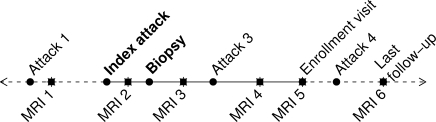
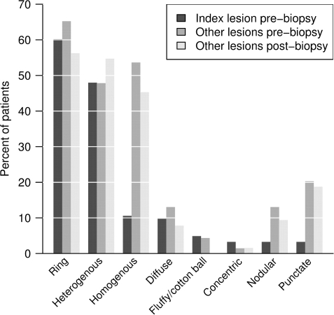
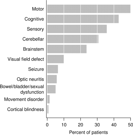
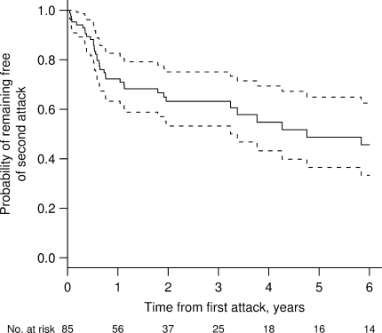
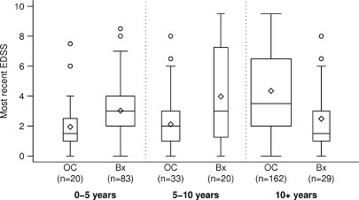
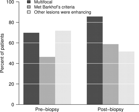
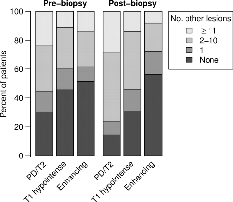
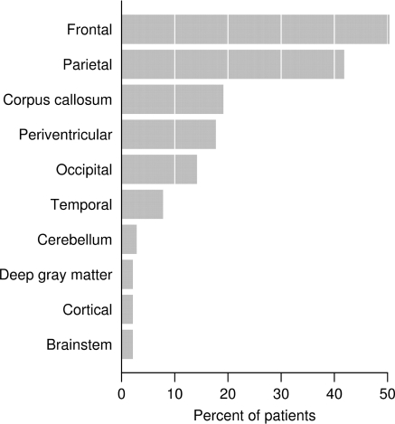
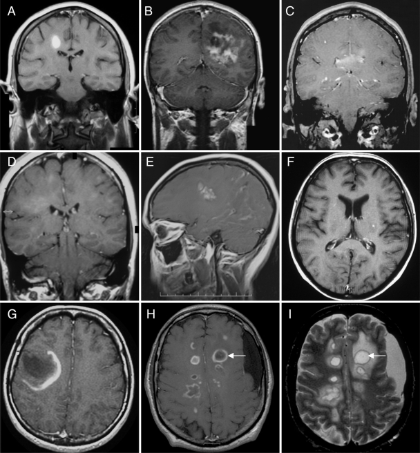
Atypical imaging features of multiple sclerosis lesions include size >2 cm, mass effect, oedema and/or ring enhancement. This constellation is often referred to as 'tumefactive multiple sclerosis'. Previous series emphasize their unifocal and clinically isolated nature, however, evolution of these lesions is not well defined. Biopsy may be required for diagnosis. We describe clinical and radiographic features in 168 patients with biopsy confirmed CNS inflammatory demyelinating disease (IDD). Lesions were analysed on pre- and post-biopsy magnetic resonance imaging (MRI) for location, size, mass effect/oedema, enhancement, multifocality and fulfilment of Barkhof criteria. Clinical data were correlated to MRI. Female to male ratio was 1.2 : 1, median age at onset, 37 years, duration between symptom onset and biopsy, 7.1 weeks and total disease duration, 3.9 years. Clinical course prior to biopsy was a first neurological event in 61%, relapsing-remitting in 29% and progressive in 4%. Presentations were typically polysymptomatic, with motor, cognitive and sensory symptoms predominating. Aphasia, agnosia, seizures and visual field defects were observed. At follow-up, 70% developed definite multiple sclerosis, and 14% had an isolated demyelinating syndrome. Median time to second attack was 4.8 years, and median EDSS at follow-up was 3.0. Multiple lesions were present in 70% on pre-biopsy MRI, and in 83% by last MRI, with Barkhof criteria fulfilled in 46% prior to biopsy and 55% by follow-up. Only 17% of cases remained unifocal. Median largest lesion size on T2-weighted images was 4 cm (range 0.5-12), with a discernible size of 2.1 cm (range 0.5-7.5). Biopsied lesions demonstrated mass effect in 45% and oedema in 77%. A strong association was found between lesion size, and presence of mass effect and/or oedema (P < 0.001). Ring enhancement was frequent. Most tumefactive features did not correlate with gender, course or diagnosis. Although lesion size >5 cm was associated with a slightly higher EDSS at last follow-up, long-term prognosis in patients with disease duration >10 years was better (EDSS 1.5) compared with a population-based multiple sclerosis cohort matched for disease duration (EDSS 3.5; P < 0.001). Given the retrospective nature of the study, the precise reason for biopsy could not always be determined. This study underscores the diagnostically challenging nature of CNS IDDs that present with atypical clinical or radiographic features. Most have multifocal disease at onset, and develop RRMS by follow-up. Although increased awareness of this broad spectrum may obviate need for biopsy in many circumstances, an important role for diagnostic brain biopsy may be required in some cases.

摘要

多发性硬化病变的非典型影像学特征包括大小>2 cm、占位效应、水肿和/或环形强化。这一组合通常被称为“瘤样多发性硬化”。既往系列研究强调其单灶性和临床孤立性,然而,这些病变的演变情况尚不明确。诊断可能需要活检。我们描述了168例经活检证实为中枢神经系统炎性脱髓鞘疾病(IDD)患者的临床和影像学特征。在活检前和活检后的磁共振成像(MRI)上分析病变的位置、大小、占位效应/水肿、强化、多灶性以及是否符合巴霍夫标准。将临床数据与MRI相关联。女性与男性比例为1.2:1,发病年龄中位数为37岁,症状出现至活检的持续时间为7.1周,疾病总持续时间为3.9年。活检前的临床病程中,61%为首次神经系统事件,29%为复发缓解型,4%为进展型。临床表现通常为多症状,以运动、认知和感觉症状为主。观察到失语、失认、癫痫发作和视野缺损。在随访中,70%发展为明确的多发性硬化,14%患有孤立性脱髓鞘综合征。第二次发作的中位时间为4.8年,随访时的扩展残疾状态量表(EDSS)中位数为3.0。活检前MRI显示70%存在多个病变,最后一次MRI显示83%存在多个病变,活检前46%符合巴霍夫标准,随访时55%符合。仅17%的病例仍为单灶性。T2加权图像上最大病变的中位大小为4 cm(范围0.5 - 12 cm),可辨别的大小为2.1 cm(范围0.5 - 7.5 cm)。活检的病变中45%显示占位效应,77%显示水肿。发现病变大小与占位效应和/或水肿的存在之间存在强关联(P < 0.)。环形强化很常见。大多数瘤样特征与性别、病程或诊断无关。尽管病变大小>5 cm与最后一次随访时略高的EDSS相关,但病程>10年患者的长期预后(EDSS 1.5)优于与之匹配的基于人群的多发性硬化队列(EDSS 3.5;P < 0.001)。鉴于该研究的回顾性性质,活检的确切原因并非总能确定。这项研究强调了具有非典型临床或影像学特征的中枢神经系统IDD在诊断上具有挑战性。大多数患者起病时为多灶性疾病,随访时发展为复发缓解型多发性硬化。尽管提高对这一广泛谱系的认识可能在许多情况下避免活检的必要性,但在某些情况下诊断性脑活检可能仍起着重要作用。

https://cdn.ncbi.nlm.nih.gov/pmc/blobs/483f/2638283/6983fc72adaa/awn098f10.jpg
https://cdn.ncbi.nlm.nih.gov/pmc/blobs/483f/2638283/ae9557033fcd/awn098f1.jpg
https://cdn.ncbi.nlm.nih.gov/pmc/blobs/483f/2638283/204f12e021e7/awn098f2.jpg
https://cdn.ncbi.nlm.nih.gov/pmc/blobs/483f/2638283/d71806df6a68/awn098f3.jpg
https://cdn.ncbi.nlm.nih.gov/pmc/blobs/483f/2638283/02146e3fa5d0/awn098f4.jpg
https://cdn.ncbi.nlm.nih.gov/pmc/blobs/483f/2638283/11d9960ed059/awn098f5.jpg
https://cdn.ncbi.nlm.nih.gov/pmc/blobs/483f/2638283/25189d94fdd3/awn098f6.jpg
https://cdn.ncbi.nlm.nih.gov/pmc/blobs/483f/2638283/59305f9a5981/awn098f7.jpg
https://cdn.ncbi.nlm.nih.gov/pmc/blobs/483f/2638283/e958d21c1f52/awn098f8.jpg
https://cdn.ncbi.nlm.nih.gov/pmc/blobs/483f/2638283/53f659969854/awn098f9.jpg
https://cdn.ncbi.nlm.nih.gov/pmc/blobs/483f/2638283/6983fc72adaa/awn098f10.jpg

相似文献

1
Clinical and radiographic spectrum of pathologically confirmed tumefactive multiple sclerosis.病理确诊的肿胀型多发性硬化症的临床和影像学表现谱
Brain. 2008 Jul;131(Pt 7):1759-75. doi: 10.1093/brain/awn098. Epub 2008 Jun 5.
2
Mass lesions in the brain: tumor or multiple sclerosis? Clinical and imaging characteristics and course from a single reference center.脑内占位性病变:肿瘤还是多发性硬化?来自单一参考中心的临床、影像学特征及病程
Turk Neurosurg. 2013;23(6):728-35. doi: 10.5137/1019-5149.JTN.7690-12.3.
3
Long-term clinical, MRI, and cognitive follow-up in a large cohort of pathologically confirmed, predominantly tumefactive multiple sclerosis.对一大群经病理证实、以肿胀型多发性硬化为主的患者进行长期临床、磁共振成像(MRI)及认知随访。
Mult Scler. 2022 Mar;28(3):441-452. doi: 10.1177/13524585211024162. Epub 2021 Jul 2.
4
Disability and T2 MRI lesions: a 20-year follow-up of patients with relapse onset of multiple sclerosis.残疾与T2加权磁共振成像病变:对复发型多发性硬化症患者的20年随访
Brain. 2008 Mar;131(Pt 3):808-17. doi: 10.1093/brain/awm329. Epub 2008 Jan 29.
5
Prognostic factors of tumefactive demyelinating lesions and differential features for multiple sclerosis in etiology.肿瘤样脱髓鞘病变的预后因素及病因学中多发性硬化的鉴别特征。
Mult Scler Relat Disord. 2024 May;85:105537. doi: 10.1016/j.msard.2024.105537. Epub 2024 Mar 5.
6
Clinical and radiological characteristics of tumefactive demyelinating lesions: follow-up study.瘤样脱髓鞘病变的临床和影像学特征:随访研究。
Mult Scler. 2012 Oct;18(10):1448-53. doi: 10.1177/1352458512438237. Epub 2012 Mar 14.
7
Is size an essential criterion to define tumefactive plaque? MR features and clinical correlation in multiple sclerosis.大小是定义瘤样斑块的必要标准吗?多发性硬化症的磁共振成像特征及临床相关性
Neuroradiol J. 2016 Oct;29(5):384-9. doi: 10.1177/1971400916665385. Epub 2016 Aug 16.
8
Inaugural tumor-like multiple sclerosis: clinical presentation and medium-term outcome in 87 patients.首例肿瘤样多发性硬化症:87 例患者的临床表现和中期预后。
J Neurol. 2018 Oct;265(10):2251-2259. doi: 10.1007/s00415-018-8984-7. Epub 2018 Jul 27.
9
Tumefactive multiple sclerosis (TMS): A case series of this challenging variant of MS.肿胀型多发性硬化症(TMS):多发性硬化症这一具有挑战性变体的病例系列。
Mult Scler Relat Disord. 2021 Feb;48:102699. doi: 10.1016/j.msard.2020.102699. Epub 2020 Dec 26.
10
Spinal cord and brain corticospinal tract lesions are associated with motor progression in tumefactive multiple sclerosis.脊髓和脑皮质脊髓束病变与肿块型多发性硬化症的运动进展有关。
Mult Scler Relat Disord. 2023 May;73:104614. doi: 10.1016/j.msard.2023.104614. Epub 2023 Mar 12.

引用本文的文献

1
Seizure as an Atypical Presentation of New-Onset Multiple Sclerosis: A Case Report.癫痫发作作为新发多发性硬化症的非典型表现:一例报告
Cureus. 2025 Mar 15;17(3):e80626. doi: 10.7759/cureus.80626. eCollection 2025 Mar.
2
Unusual Stroke-Like Presentation of Tumefactive Multiple Sclerosis in a 27-Year-Old Man.一名27岁男性的肿胀型多发性硬化症的不寻常卒中样表现
Cureus. 2025 Mar 2;17(3):e79909. doi: 10.7759/cureus.79909. eCollection 2025 Mar.
3
Mood Disorder and Multimorbidity Complicating a Multiple Sclerosis Diagnosis: From the National Multiple Sclerosis Society Case Conference Proceedings.

本文引用的文献

1
Pitfalls in the diagnosis of brain tumours.脑肿瘤诊断中的陷阱。
Lancet Neurol. 2006 Nov;5(11):937-48. doi: 10.1016/S1474-4422(06)70597-X.
2
MRI and the diagnosis of multiple sclerosis: expanding the concept of "no better explanation".磁共振成像与多发性硬化症的诊断:拓展“无更好解释”的概念
Lancet Neurol. 2006 Oct;5(10):841-52. doi: 10.1016/S1474-4422(06)70572-5.
3
Multiple sclerosis, brain radiotherapy, and risk of neurotoxicity: the Mayo Clinic experience.多发性硬化症、脑部放射治疗与神经毒性风险:梅奥诊所的经验
情绪障碍与多种疾病并存使多发性硬化症诊断复杂化:来自美国国家多发性硬化症协会病例会议记录
Neurol Neuroimmunol Neuroinflamm. 2025 Mar;12(2):e200376. doi: 10.1212/NXI.0000000000200376. Epub 2025 Feb 11.
4
CNS B cell infiltration in tumefactive anti-myelin oligodendrocyte glycoprotein antibody-associated disease.肿胀性抗髓鞘少突胶质细胞糖蛋白抗体相关疾病中的中枢神经系统B细胞浸润
Mult Scler J Exp Transl Clin. 2024 Dec 5;10(4):20552173241301011. doi: 10.1177/20552173241301011. eCollection 2024 Oct-Dec.
5
The diagnostic challenge of differentiating tumefactive multiple sclerosis (TMS) from other brain lesions: a case report and literature review on a rare subtype of MS.鉴别肿胀型多发性硬化(TMS)与其他脑部病变的诊断挑战:一例病例报告及关于多发性硬化罕见亚型的文献综述
Ann Med Surg (Lond). 2024 Oct 24;86(12):7418-7422. doi: 10.1097/MS9.0000000000002687. eCollection 2024 Dec.
6
Establishment of an MRI-based radiomics model for distinguishing between intramedullary spinal cord tumor and tumefactive demyelinating lesion.建立基于 MRI 的放射组学模型以区分脊髓髓内肿瘤和肿块样脱髓鞘病变。
BMC Med Imaging. 2024 Nov 21;24(1):317. doi: 10.1186/s12880-024-01499-8.
7
Subcortical plaques and inflammation reflect cortical and meningeal pathologies in progressive multiple sclerosis.皮质下斑块和炎症反映了进行性多发性硬化症中的皮质和脑膜病变。
Brain Pathol. 2025 May;35(3):e13314. doi: 10.1111/bpa.13314. Epub 2024 Oct 26.
8
Long-term outcomes of ADEM-like and tumefactive presentations of CNS demyelination: a case-comparison analysis.CNS 脱髓鞘疾病中 ADEM 样和肿块型表现的长期预后:病例对照分析。
J Neurol. 2024 Aug;271(8):5275-5289. doi: 10.1007/s00415-024-12349-6. Epub 2024 Jun 11.
9
A Rare and Challenging Presentation of Acute Hemorrhagic Leukoencephalitis With Tumefactive Demyelinating Lesions in a 41-Year-Old Male.一名41岁男性急性出血性白质脑炎合并瘤样脱髓鞘病变的罕见且具有挑战性的病例表现
Cureus. 2024 Apr 15;16(4):e58282. doi: 10.7759/cureus.58282. eCollection 2024 Apr.
10
Tumor-like Lesions in Primary Angiitis of the Central Nervous System: The Role of Magnetic Resonance Imaging in Differential Diagnosis.中枢神经系统原发性血管炎中的肿瘤样病变:磁共振成像在鉴别诊断中的作用
Diagnostics (Basel). 2024 Mar 14;14(6):618. doi: 10.3390/diagnostics14060618.
Int J Radiat Oncol Biol Phys. 2006 Nov 15;66(4):1178-86. doi: 10.1016/j.ijrobp.2006.06.014. Epub 2006 Sep 11.
4
Absence of cerebrospinal fluid oligoclonal bands is associated with delayed disability progression in relapsing-remitting MS patients treated with interferon-beta.在接受β-干扰素治疗的复发缓解型多发性硬化症患者中,脑脊液寡克隆带缺失与残疾进展延迟相关。
J Neurol Sci. 2006 May 15;244(1-2):97-102. doi: 10.1016/j.jns.2006.01.004. Epub 2006 Feb 13.
5
Pattern of T2 hypointensity associated with ring-enhancing brain lesions can help to differentiate pathology.与脑环形强化病变相关的T2低信号模式有助于鉴别病理情况。
Neuroradiology. 2006 Mar;48(3):143-9. doi: 10.1007/s00234-005-0024-5. Epub 2006 Jan 31.
6
Clinical course, pathological correlations, and outcome of biopsy proved inflammatory demyelinating disease.活检证实的炎性脱髓鞘疾病的临床病程、病理关联及转归
J Neurol Neurosurg Psychiatry. 2005 Dec;76(12):1693-7. doi: 10.1136/jnnp.2004.060624.
7
Tumefactive demyelinating lesions: conventional and advanced magnetic resonance imaging.肿块样脱髓鞘病变:传统及先进的磁共振成像
Mult Scler. 2005 Apr;11(2):135-9. doi: 10.1191/1352458505ms1145oa.
8
Multiple sclerosis: pseudotumoral forms.多发性硬化症:假瘤样形式。
Neurol Sci. 2004 Nov;25 Suppl 4:S374-9. doi: 10.1007/s10072-004-0345-x.
9
Late-onset multiple sclerosis mimicking brain tumor: a case report.酷似脑肿瘤的迟发性多发性硬化症:一例报告
Brain Tumor Pathol. 2004;21(2):83-6. doi: 10.1007/BF02484515.
10
Role of magnetic resonance imaging within diagnostic criteria for multiple sclerosis.磁共振成像在多发性硬化诊断标准中的作用。
Ann Neurol. 2004 Aug;56(2):273-8. doi: 10.1002/ana.20156.