López María R, Schlegel Elisabeth F M, Wintersteller Sandra, Blaho John A
Department of Microbiology, Mount Sinai School of Medicine, One Gustave L. Levy, New York, NY 10029, USA.
Virus Res. 2008 Sep;136(1-2):175-88. doi: 10.1016/j.virusres.2008.05.010. Epub 2008 Jun 12.
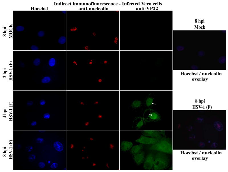
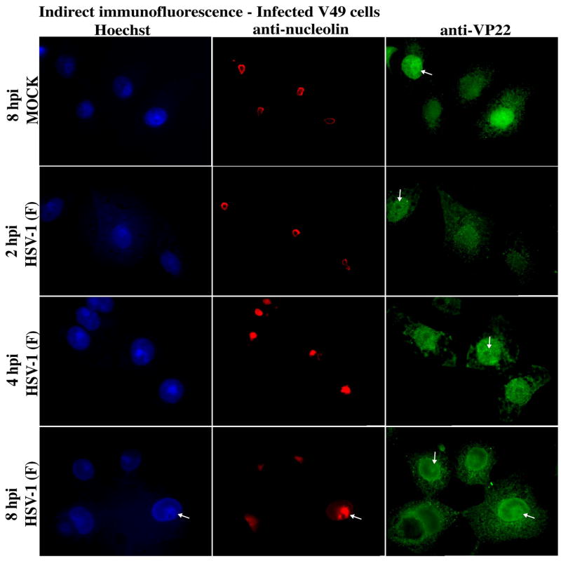
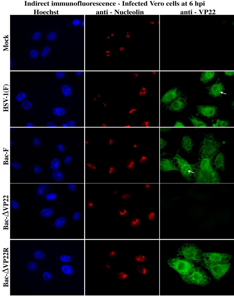
The herpes simplex virus (HSV) major tegument structural protein VP22 resides in multiple subcellular regions during productive infection. During an analysis of the molecular determinants of these localizations, we observed that a transfected fusion of the C-terminal portion of VP22, containing its pat4 nuclear localization signal, with GFP lacked nucleolar sparing compared to GFP alone. Thus, the initial goal was to determine whether VP22 associates with nucleoli. Using an optimized indirect immunofluorescence system to visualize nucleolin and viral proteins, we observed that VP22 present in VP22-expressing Vero (V49) cells "surrounded" nucleolin. These two initial findings implied that VP22 might associate directly with nucleoli. We next analyzed HSV-infected cells and observed that at late times, anti-nucleolin immune reactivity was dispersed throughout the nuclei while it retained uniform, circular staining in mock-infected cells. Time course infection experiments indicated that nucleolin initiated its transition from uniform to dispersed structures between 2 and 4 hpi. Comparison of Hoechst stained nuclei showed bright anti-nucleolin staining localized to regions of marginalized chromatin. These effects required de novo infected cell protein synthesis. A portion of VP22 detected in nuclei at 4 and 6 hpi localized to these areas of altered nucleolin and marginalized chromatin. VP22 was excluded from viral replication compartments containing the viral regulatory protein ICP22. Finally, altered nucleolin and marginalized chromatin were detected with a VP22-null virus, indicating that VP22 was not responsible for these nuclear architecture alterations. Thus, we conclude that nuclear VP22 targets unique subnuclear structures early (<6hpi) during herpes simplex virus 1 (HSV-1) infection.
单纯疱疹病毒(HSV)主要被膜结构蛋白VP22在增殖性感染期间定位于多个亚细胞区域。在对这些定位的分子决定因素进行分析的过程中,我们观察到,与单独的绿色荧光蛋白(GFP)相比,转染的包含其pat4核定位信号的VP22 C末端部分与GFP的融合蛋白缺乏核仁保留现象。因此,最初的目标是确定VP22是否与核仁相关。使用优化的间接免疫荧光系统来可视化核仁素和病毒蛋白,我们观察到在表达VP22的非洲绿猴肾(V49)细胞中存在的VP22 “包围” 了核仁素。这两个初步发现表明VP22可能直接与核仁相关。接下来,我们分析了HSV感染的细胞,观察到在感染后期,抗核仁素免疫反应性分散在整个细胞核中,而在 mock 感染的细胞中它保持均匀的圆形染色。时间进程感染实验表明,核仁素在感染后2至4小时之间开始从均匀结构转变为分散结构。Hoechst染色细胞核的比较显示,明亮的抗核仁素染色定位于边缘化染色质区域。这些效应需要从头进行感染细胞的蛋白质合成。在感染后4小时和6小时在细胞核中检测到的一部分VP22定位于核仁素改变和边缘化染色质的这些区域。VP22被排除在含有病毒调节蛋白ICP22的病毒复制区室之外。最后,用VP22缺失病毒检测到了核仁素改变和边缘化染色质,这表明VP22与这些核结构改变无关。因此,我们得出结论,在单纯疱疹病毒1(HSV-1)感染期间,核VP22在早期(<6小时)靶向独特的亚核结构。