Babon Jeffrey J, Sabo Jennifer K, Soetopo Alfreda, Yao Shenggen, Bailey Michael F, Zhang Jian-Guo, Nicola Nicos A, Norton Raymond S
Walter and Eliza Hall Institute of Medical Research, 1G Royal Parade, Parkville, Victoria 3050, Australia.
J Mol Biol. 2008 Sep 12;381(4):928-40. doi: 10.1016/j.jmb.2008.06.038. Epub 2008 Jun 20.
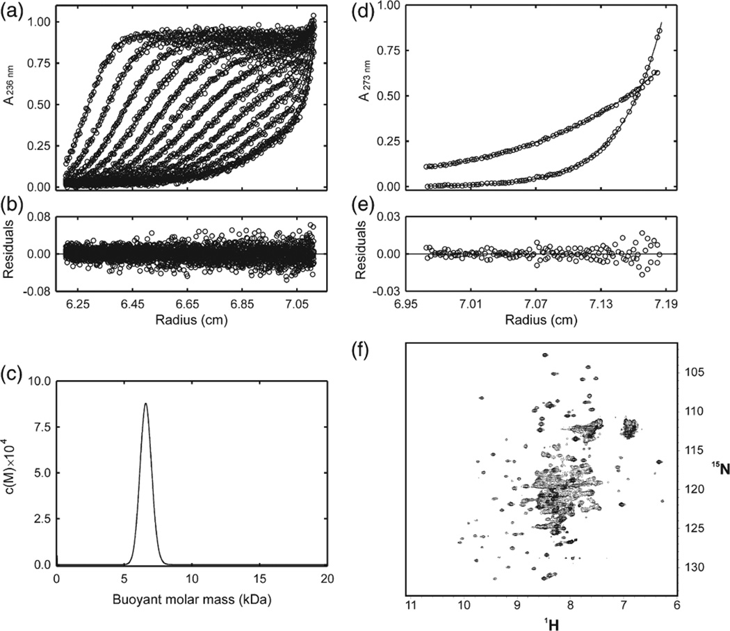
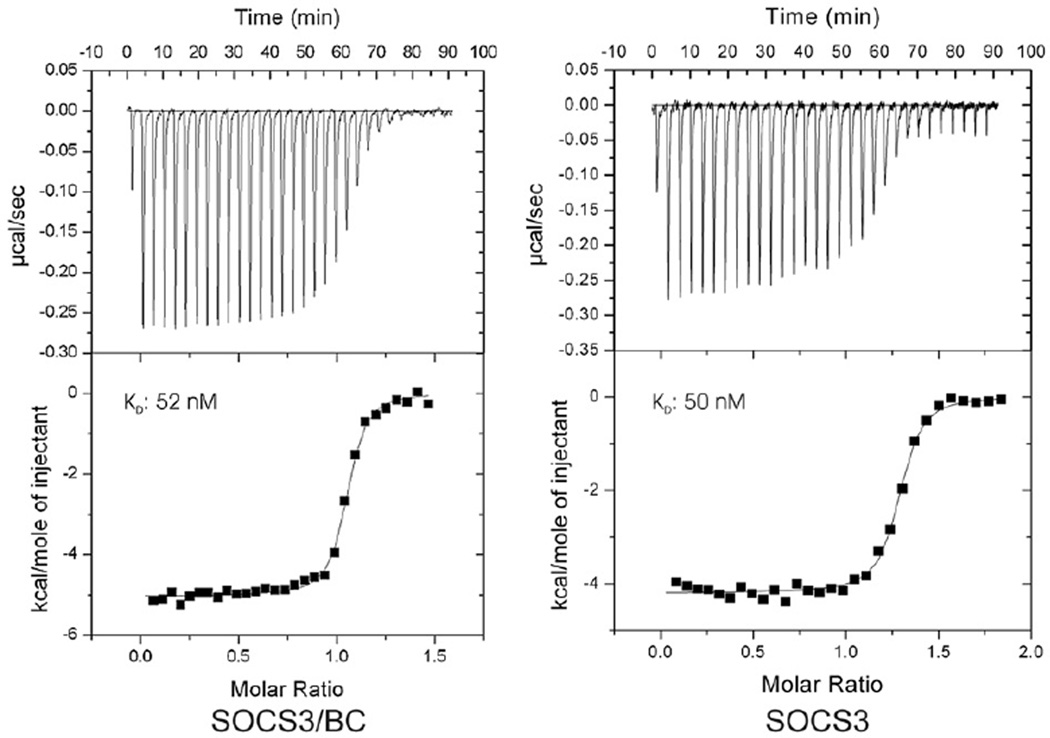
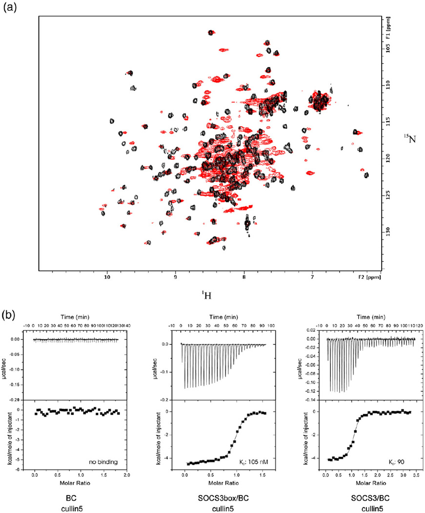
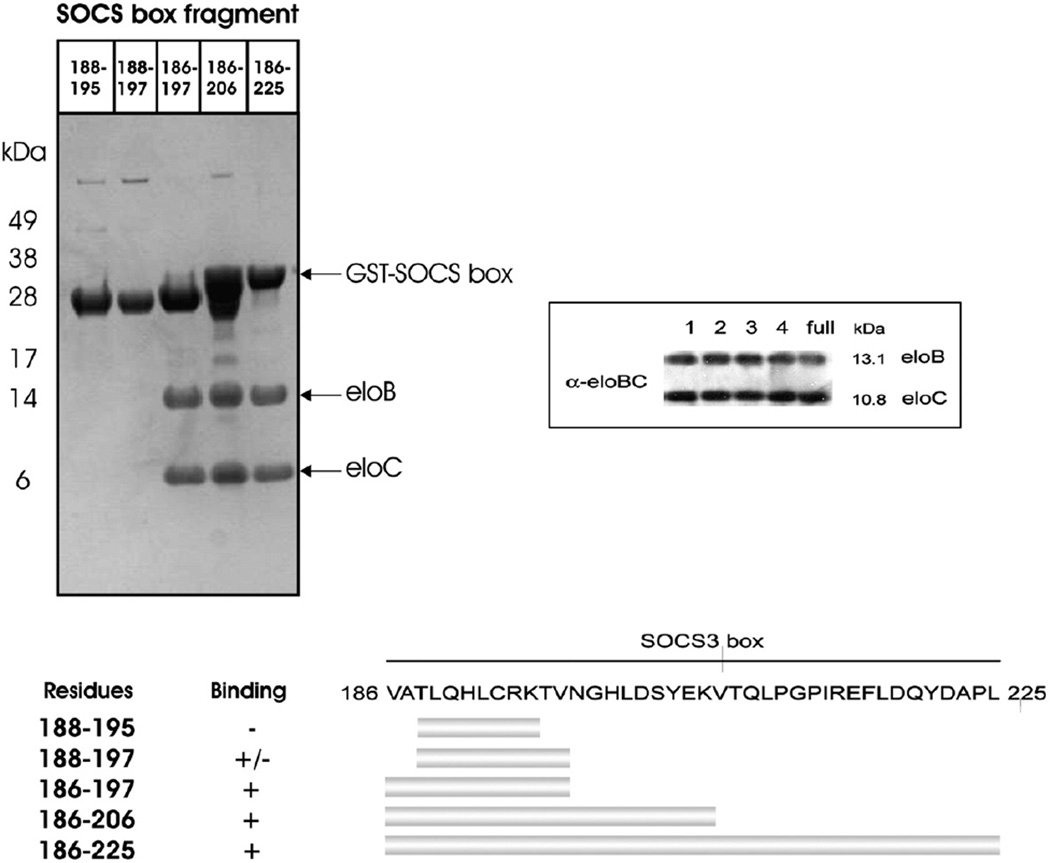
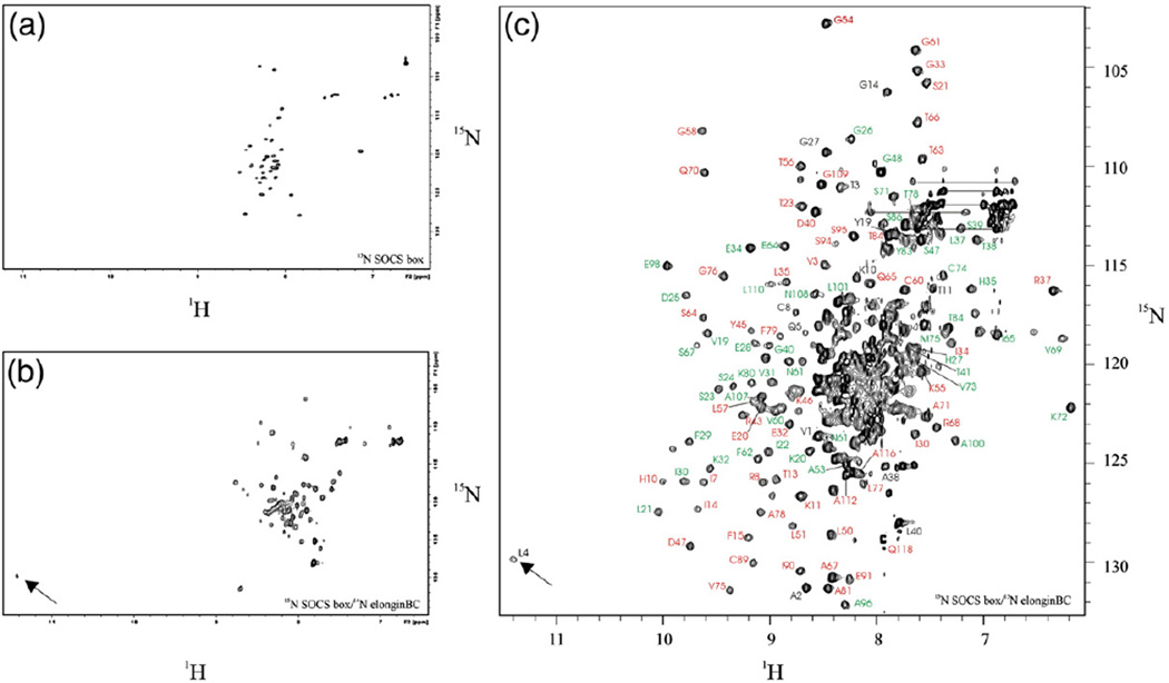
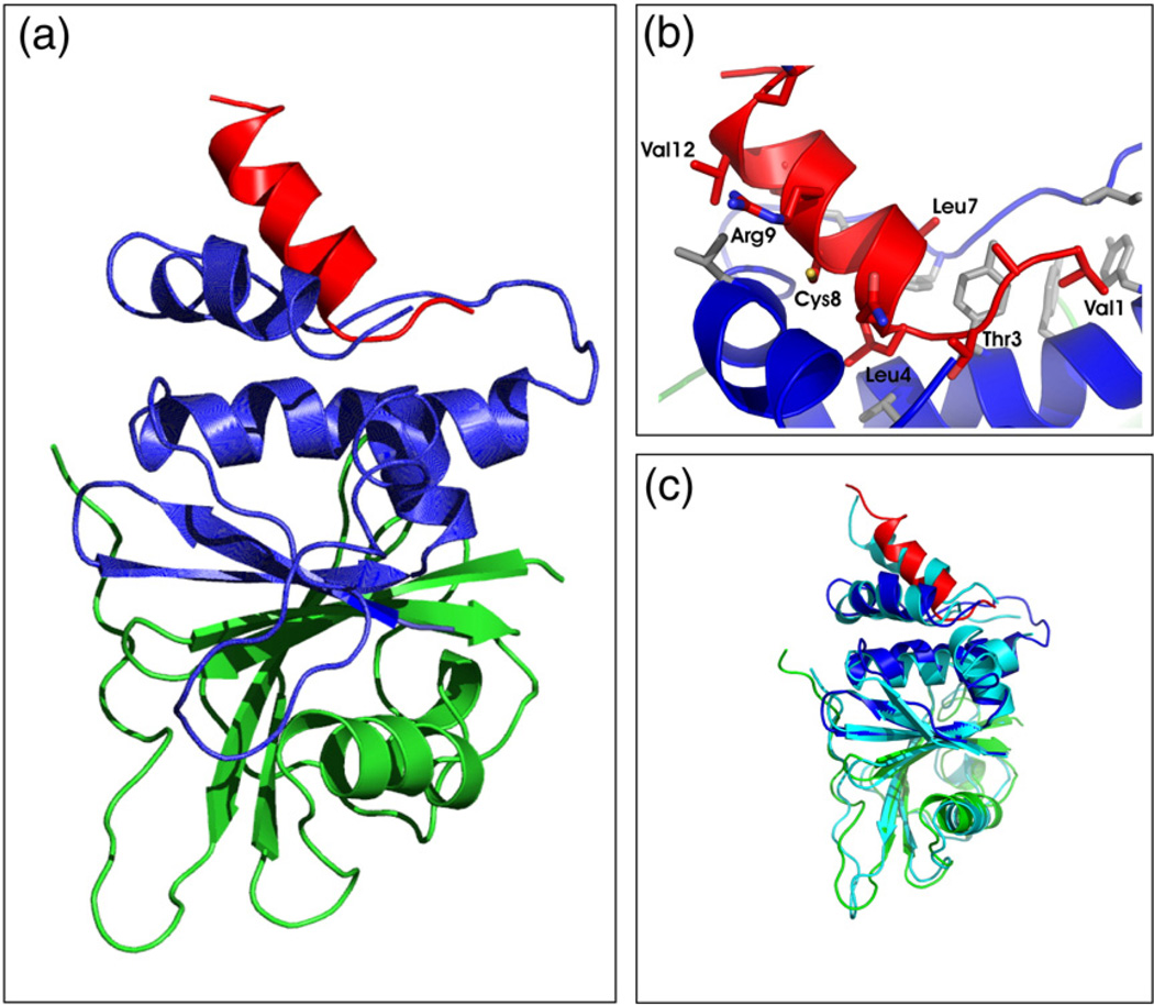
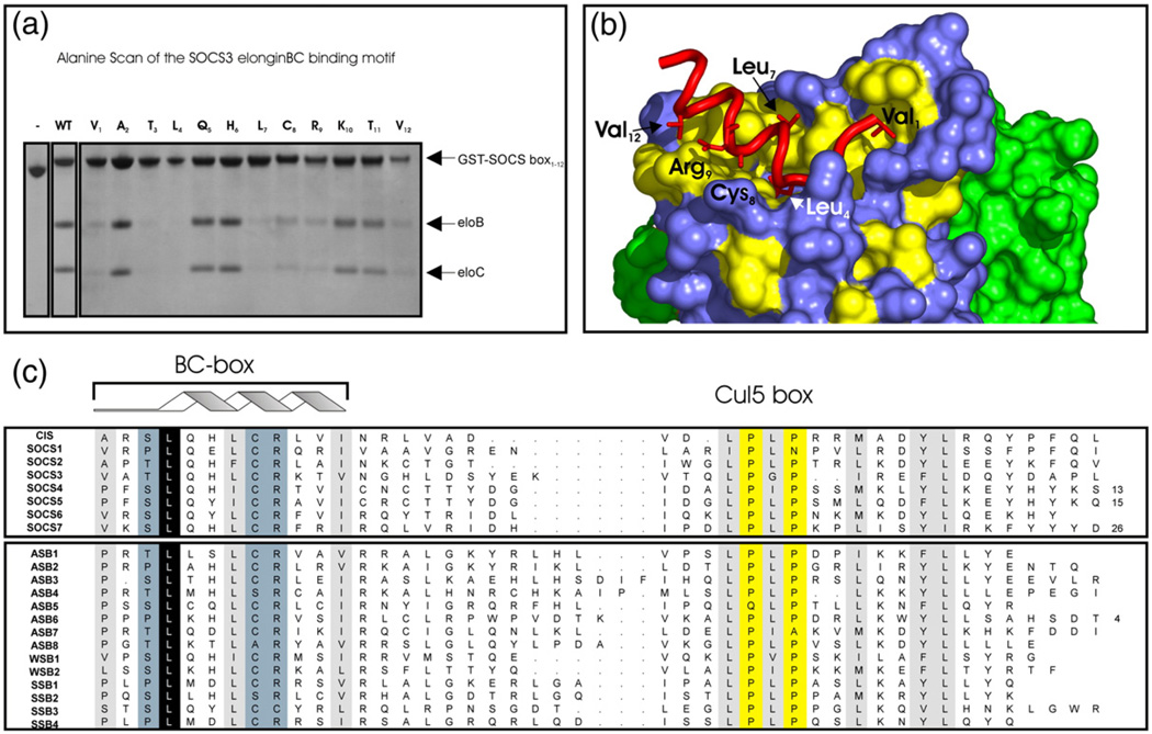
Suppressor of cytokine signalling 3 (SOCS3) is responsible for regulating the cellular response to a variety of cytokines, including interleukin 6 and leukaemia inhibitory factor. Identification of the SOCS box domain led to the hypothesis that SOCS3 can associate with functional E3 ubiquitin ligases and thereby induce the degradation of bound signalling proteins. This model relies upon an interaction between the SOCS box, elonginBC and a cullin protein that forms the E3 ligase scaffold. We have investigated this interaction in vitro using purified components and show that SOCS3 binds to elonginBC and cullin5 with high affinity. The SOCS3-elonginBC interaction was further characterised by determining the solution structure of the SOCS box-elonginBC ternary complex and by deletion and alanine scanning mutagenesis of the SOCS box. These studies revealed that conformational flexibility is a key feature of the SOCS-elonginBC interaction. In particular, the SOCS box is disordered in isolation and only becomes structured upon elonginBC association. The interaction depends upon the first 12 residues of the SOCS box domain and particularly on a deeply buried, conserved leucine. The SOCS box, when bound to elonginBC, binds tightly to cullin5 with 100 nM affinity. Domains upstream of the SOCS box are not required for elonginBC or cullin5 association, indicating that the SOCS box acts as an independent binding domain capable of recruiting elonginBC and cullin5 to promote E3 ligase formation.
细胞因子信号转导抑制因子3(SOCS3)负责调节细胞对多种细胞因子的反应,包括白细胞介素6和白血病抑制因子。SOCS框结构域的鉴定引发了一种假说,即SOCS3可与功能性E3泛素连接酶结合,从而诱导结合的信号蛋白降解。该模型依赖于SOCS框、延伸蛋白BC和形成E3连接酶支架的cullin蛋白之间的相互作用。我们使用纯化的组分在体外研究了这种相互作用,结果表明SOCS3以高亲和力与延伸蛋白BC和cullin5结合。通过确定SOCS框-延伸蛋白BC三元复合物的溶液结构以及对SOCS框进行缺失和丙氨酸扫描诱变,进一步对SOCS3-延伸蛋白BC相互作用进行了表征。这些研究表明,构象灵活性是SOCS-延伸蛋白BC相互作用的关键特征。特别是,SOCS框单独存在时无序,仅在与延伸蛋白BC结合后才形成结构。这种相互作用取决于SOCS框结构域的前12个残基,尤其依赖于一个深埋的保守亮氨酸。当与延伸蛋白BC结合时,SOCS框以100 nM的亲和力紧密结合cullin5。SOCS框上游的结构域对于与延伸蛋白BC或cullin5的结合不是必需的,这表明SOCS框作为一个独立的结合结构域,能够募集延伸蛋白BC和cullin5以促进E3连接酶的形成。