Vaid Moninder, Claydon Thomas W, Rezazadeh Saman, Fedida David
Department of Anesthesiology, Pharmacology, and Therapeutics, University of British Columbia, Vancouver, BC V6T 1Z3, Canada.
J Gen Physiol. 2008 Aug;132(2):209-22. doi: 10.1085/jgp.200809978. Epub 2008 Jul 14.
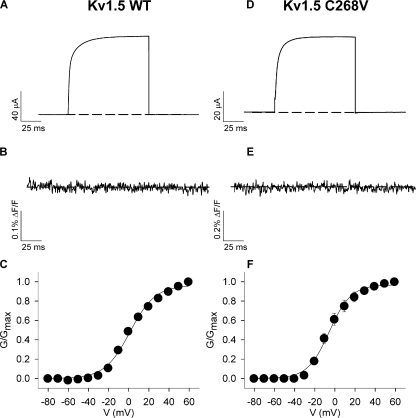
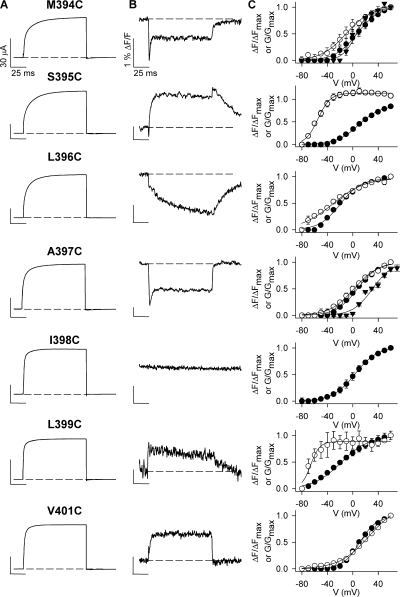
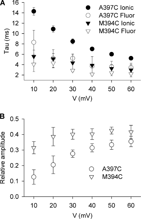
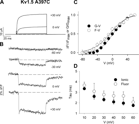
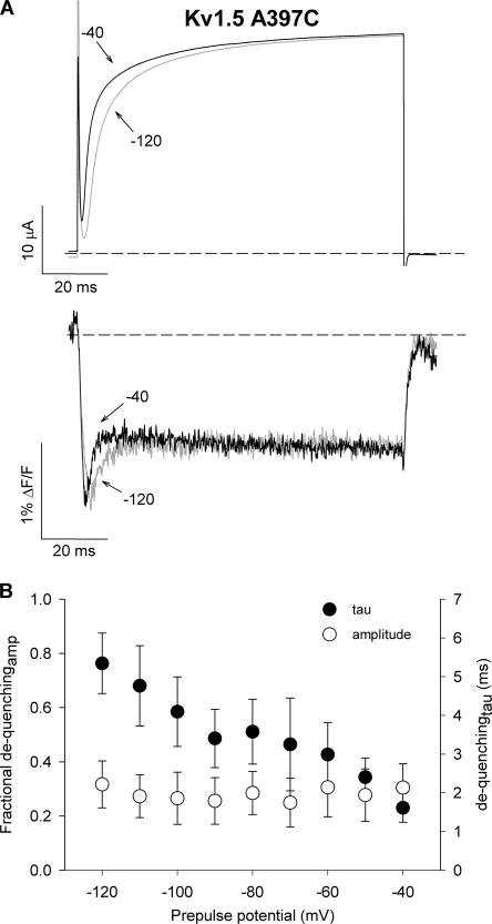
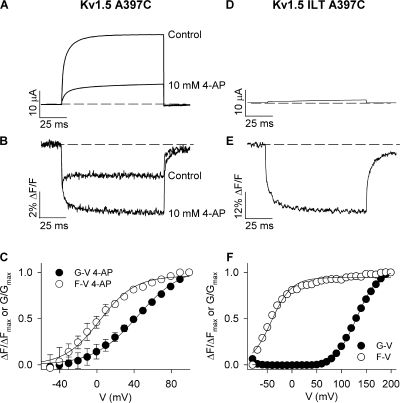
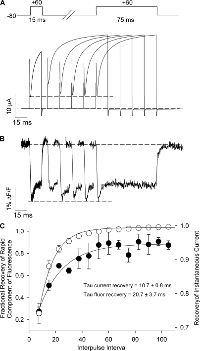
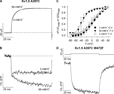
Voltage-gated potassium (Kv) channel gating involves complex structural rearrangements that regulate the ability of channels to conduct K(+) ions. Fluorescence-based approaches provide a powerful technique to directly report structural dynamics underlying these gating processes in Shaker Kv channels. Here, we apply voltage clamp fluorimetry, for the first time, to study voltage sensor motions in mammalian Kv1.5 channels. Despite the homology between Kv1.5 and the Shaker channel, attaching TMRM or PyMPO fluorescent probes to substituted cysteine residues in the S3-S4 linker of Kv1.5 (M394C-V401C) revealed unique and unusual fluorescence signals. Whereas the fluorescence during voltage sensor movement in Shaker channels was monoexponential and occurred with a similar time course to ionic current activation, the fluorescence report of Kv1.5 voltage sensor motions was transient with a prominent rapidly dequenching component that, with TMRM at A397C (equivalent to Shaker A359C), represented 36 +/- 3% of the total signal and occurred with a tau of 3.4 +/- 0.6 ms at +60 mV (n = 4). Using a number of approaches, including 4-AP drug block and the ILT triple mutation, which dissociate channel opening from voltage sensor movement, we demonstrate that the unique dequenching component of fluorescence is associated with channel opening. By regulating the outer pore structure using raised (99 mM) external K(+) to stabilize the conducting configuration of the selectivity filter, or the mutations W472F (equivalent to Shaker W434F) and H463G to stabilize the nonconducting (P-type inactivated) configuration of the selectivity filter, we show that the dequenching of fluorescence reflects rapid structural events at the selectivity filter gate rather than the intracellular pore gate.
电压门控钾(Kv)通道的门控涉及复杂的结构重排,这些重排调节通道传导K⁺离子的能力。基于荧光的方法提供了一种强大的技术,可直接报告Shaker Kv通道中这些门控过程背后的结构动力学。在这里,我们首次应用电压钳荧光法来研究哺乳动物Kv1.5通道中的电压传感器运动。尽管Kv1.5与Shaker通道具有同源性,但将TMRM或PyMPO荧光探针连接到Kv1.5(M394C-V401C)的S3-S4连接子中的取代半胱氨酸残基上,却揭示了独特且不寻常的荧光信号。在Shaker通道中,电压传感器运动期间的荧光是单指数的,并且其发生的时间进程与离子电流激活相似,而Kv1.5电压传感器运动的荧光报告是瞬态的,具有一个突出的快速去淬灭成分,在A397C(相当于Shaker A359C)处使用TMRM时,该成分占总信号的36±3%,在+60 mV时的时间常数为3.4±0.6 ms(n = 4)。使用多种方法,包括4-AP药物阻断和ILT三重突变,这些方法可使通道开放与电压传感器运动解离,我们证明荧光的独特去淬灭成分与通道开放有关。通过使用升高的(99 mM)外部K⁺调节外孔结构以稳定选择性过滤器的传导构型,或使用突变W472F(相当于Shaker W434F)和H463G来稳定选择性过滤器的非传导(P型失活)构型,我们表明荧光的去淬灭反映了选择性过滤器门处的快速结构事件,而不是细胞内孔门处的事件。