• 文献检索
  • 文档翻译
  • 深度研究
  • 学术资讯
  • Suppr Zotero 插件Zotero 插件
  • 邀请有礼
  • 套餐&价格
  • 历史记录
应用&插件
Suppr Zotero 插件Zotero 插件浏览器插件Mac 客户端Windows 客户端微信小程序
定价
高级版会员购买积分包购买API积分包
服务
文献检索文档翻译深度研究API 文档MCP 服务
关于我们
关于 Suppr公司介绍联系我们用户协议隐私条款
关注我们

Suppr 超能文献

核心技术专利:CN118964589B侵权必究
粤ICP备2023148730 号-1Suppr @ 2026

文献检索

告别复杂PubMed语法,用中文像聊天一样搜索,搜遍4000万医学文献。AI智能推荐,让科研检索更轻松。

立即免费搜索

文件翻译

保留排版,准确专业,支持PDF/Word/PPT等文件格式,支持 12+语言互译。

免费翻译文档

深度研究

AI帮你快速写综述,25分钟生成高质量综述,智能提取关键信息,辅助科研写作。

立即免费体验

三维空间关系分析:神经细胞模式研究工具

Analysis of spatial relationships in three dimensions: tools for the study of nerve cell patterning.

作者信息

Eglen Stephen J, Lofgreen Dan D, Raven Mary A, Reese Benjamin E

机构信息

Cambridge Computational Biology Institute, Department of Applied Mathematics and Theoretical Physics, University of Cambridge, Cambridge, CB3 0WA, UK.

出版信息

BMC Neurosci. 2008 Jul 21;9:68. doi: 10.1186/1471-2202-9-68.

DOI:10.1186/1471-2202-9-68
PMID:18644136
原文链接:https://pmc.ncbi.nlm.nih.gov/articles/PMC2491625/
Abstract

BACKGROUND

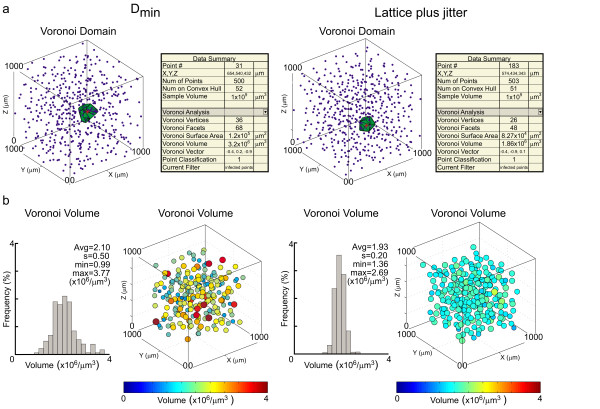
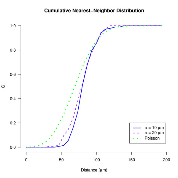
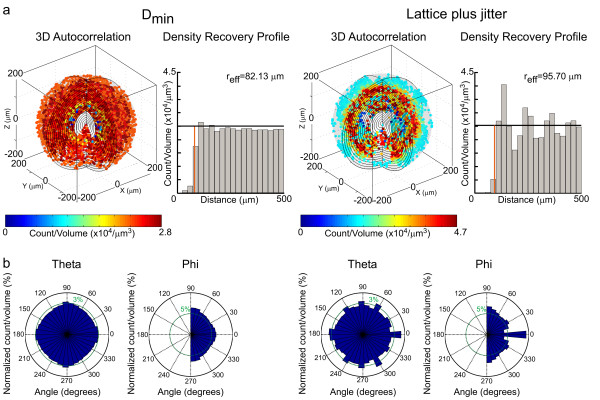
Multiple technologies have been brought to bear on understanding the three-dimensional morphology of individual neurons and glia within the brain, but little progress has been made on understanding the rules controlling cellular patterning. We describe new matlab-based software tools, now available to the scientific community, permitting the calculation of spatial statistics associated with 3D point patterns. The analyses are largely derived from the Delaunay tessellation of the field, including the nearest neighbor and Voronoi domain analyses, and from the spatial autocorrelogram.

RESULTS

Our tools enable the analysis of the spatial relationship between neurons within the central nervous system in 3D, and permit the modeling of these fields based on lattice-like simulations, and on simulations of minimal-distance spacing rules. Here we demonstrate the utility of our analysis methods to discriminate between two different simulated neuronal populations.

CONCLUSION

Together, these tools can be used to reveal the presence of nerve cell patterning and to model its foundation, in turn informing on the potential developmental mechanisms that govern its establishment. Furthermore, in conjunction with analyses of dendritic morphology, they can be used to determine the degree of dendritic coverage within a volume of tissue exhibited by mature nerve cells.

摘要

背景

多种技术已被用于理解大脑中单个神经元和神经胶质细胞的三维形态,但在理解控制细胞模式形成的规则方面进展甚微。我们描述了一种基于Matlab的新软件工具,现已向科学界提供,它可以计算与三维点模式相关的空间统计量。这些分析主要源于该区域的德劳内三角剖分,包括最近邻分析和沃罗诺伊域分析,以及空间自相关图。

结果

我们的工具能够对中枢神经系统中神经元之间的空间关系进行三维分析,并允许基于类似晶格的模拟以及最小距离间隔规则的模拟对这些区域进行建模。在此,我们展示了我们的分析方法在区分两种不同模拟神经元群体方面的效用。

结论

总之,这些工具可用于揭示神经细胞模式的存在并对其基础进行建模,进而为控制其形成的潜在发育机制提供信息。此外,结合树突形态分析,它们可用于确定成熟神经细胞在一定体积组织内的树突覆盖程度。

https://cdn.ncbi.nlm.nih.gov/pmc/blobs/602c/2491625/a7f391631b98/1471-2202-9-68-3.jpg
https://cdn.ncbi.nlm.nih.gov/pmc/blobs/602c/2491625/d8d1c62365bb/1471-2202-9-68-1.jpg
https://cdn.ncbi.nlm.nih.gov/pmc/blobs/602c/2491625/5175c019cecd/1471-2202-9-68-2.jpg
https://cdn.ncbi.nlm.nih.gov/pmc/blobs/602c/2491625/a7f391631b98/1471-2202-9-68-3.jpg
https://cdn.ncbi.nlm.nih.gov/pmc/blobs/602c/2491625/d8d1c62365bb/1471-2202-9-68-1.jpg
https://cdn.ncbi.nlm.nih.gov/pmc/blobs/602c/2491625/5175c019cecd/1471-2202-9-68-2.jpg
https://cdn.ncbi.nlm.nih.gov/pmc/blobs/602c/2491625/a7f391631b98/1471-2202-9-68-3.jpg

相似文献

1
Analysis of spatial relationships in three dimensions: tools for the study of nerve cell patterning.三维空间关系分析:神经细胞模式研究工具
BMC Neurosci. 2008 Jul 21;9:68. doi: 10.1186/1471-2202-9-68.
2
Ka-me: a Voronoi image analyzer.卡美:一种 Voronoi 图像分析器。
Bioinformatics. 2012 Jul 1;28(13):1802-4. doi: 10.1093/bioinformatics/bts253. Epub 2012 May 3.
3
Modular approach for resolving and mapping complex neural and other cellular structures and their associated deformation fields in three dimensions.用于解析和映射三维复杂神经和其他细胞结构及其相关变形场的模块化方法。
Nat Protoc. 2018 Dec;13(12):3042-3064. doi: 10.1038/s41596-018-0077-7.
4
A Quantitative Three-Dimensional Image Analysis Tool for Maximal Acquisition of Spatial Heterogeneity Data.一种用于最大程度获取空间异质性数据的定量三维图像分析工具。
Tissue Eng Part C Methods. 2017 Feb;23(2):108-117. doi: 10.1089/ten.TEC.2016.0413.
5
Cloud based toolbox for image analysis, processing and reconstruction tasks.用于图像分析、处理和重建任务的基于云的工具箱。
Adv Exp Med Biol. 2015;823:191-205. doi: 10.1007/978-3-319-10984-8_11.
6
Neuron tracing in perspective.神经示踪技术的透视。
Cytometry A. 2010 Jul;77(7):693-704. doi: 10.1002/cyto.a.20895.
7
Modelling the magnetic signature of neuronal tissue.模拟神经组织的磁信号特征。
Neuroimage. 2007 Aug 1;37(1):137-48. doi: 10.1016/j.neuroimage.2007.04.033. Epub 2007 May 4.
8
VAST (Volume Annotation and Segmentation Tool): Efficient Manual and Semi-Automatic Labeling of Large 3D Image Stacks.VAST(体积注释和分割工具):高效的大型 3D 图像堆栈手动和半自动标注。
Front Neural Circuits. 2018 Oct 16;12:88. doi: 10.3389/fncir.2018.00088. eCollection 2018.
9
Proof-editing is the bottleneck of 3D neuron reconstruction: the problem and solutions.校对编辑是3D神经元重建的瓶颈:问题与解决方案
Neuroinformatics. 2011 Sep;9(2-3):103-5. doi: 10.1007/s12021-010-9090-x.
10
Imaging cellular network dynamics in three dimensions using fast 3D laser scanning.使用快速三维激光扫描对细胞网络动力学进行三维成像。
Nat Methods. 2007 Jan;4(1):73-9. doi: 10.1038/nmeth989. Epub 2006 Dec 3.

引用本文的文献

1
CytoSpatio: Learning cell type spatial relationships using multirange, multitype point process models.细胞空间分析:使用多范围、多类型点过程模型学习细胞类型空间关系。
PLoS Comput Biol. 2025 Aug 21;21(8):e1013409. doi: 10.1371/journal.pcbi.1013409. eCollection 2025 Aug.
2
CytoSpatio: Learning cell type spatial relationships using multirange, multitype point process models.细胞空间分析:使用多范围、多类型点过程模型学习细胞类型空间关系。
bioRxiv. 2024 Nov 3:2024.10.31.621408. doi: 10.1101/2024.10.31.621408.
3
Quantitative 3D investigation of Neuronal network in mouse spinal cord model.

本文引用的文献

1
Novel applications of common stereology software to represent the complete distribution, density and spatial organization of anterogradely labelled fibers in neuroanatomical tract-tracing studies.
J Neurosci Methods. 2007 Jun 15;163(1):17-23. doi: 10.1016/j.jneumeth.2007.02.005. Epub 2007 Feb 15.
2
Development of regular cellular spacing in the retina: theoretical models.视网膜中规则细胞间距的发育:理论模型
Math Med Biol. 2006 Jun;23(2):79-99. doi: 10.1093/imammb/dql003. Epub 2006 Mar 1.
3
Developmental improvement in the regularity and packing of mouse horizontal cells: implications for mechanisms underlying mosaic pattern formation.小鼠水平细胞规则性和排列的发育改善:对镶嵌模式形成潜在机制的启示
定量 3D 研究小鼠脊髓模型中的神经元网络。
Sci Rep. 2017 Jan 23;7:41054. doi: 10.1038/srep41054.
4
Quantitative Imaging of Cholinergic Interneurons Reveals a Distinctive Spatial Organization and a Functional Gradient across the Mouse Striatum.胆碱能中间神经元的定量成像揭示了小鼠纹状体内独特的空间组织和功能梯度。
PLoS One. 2016 Jun 17;11(6):e0157682. doi: 10.1371/journal.pone.0157682. eCollection 2016.
5
Topological analyses in APP/PS1 mice reveal that astrocytes do not migrate to amyloid-β plaques.对APP/PS1小鼠进行的拓扑分析表明,星形胶质细胞不会迁移至β淀粉样蛋白斑块处。
Proc Natl Acad Sci U S A. 2015 Dec 22;112(51):15556-61. doi: 10.1073/pnas.1516779112. Epub 2015 Dec 7.
6
Editorial: Quantitative Analysis of Neuroanatomy.社论:神经解剖学的定量分析
Front Neuroanat. 2015 Nov 11;9:143. doi: 10.3389/fnana.2015.00143. eCollection 2015.
7
Design principles and developmental mechanisms underlying retinal mosaics.视网膜镶嵌的设计原则和发育机制。
Biol Rev Camb Philos Soc. 2015 Aug;90(3):854-76. doi: 10.1111/brv.12139. Epub 2014 Aug 8.
8
Spatial distribution of neurons innervated by chandelier cells.由吊灯细胞支配的神经元的空间分布。
Brain Struct Funct. 2015 Sep;220(5):2817-34. doi: 10.1007/s00429-014-0828-3. Epub 2014 Jul 24.
9
RipleyGUI: software for analyzing spatial patterns in 3D cell distributions.RipleyGUI:用于分析 3D 细胞分布空间模式的软件。
Front Neuroinform. 2013 Apr 9;7:5. doi: 10.3389/fninf.2013.00005. eCollection 2013.
10
Three-dimensional spatial distribution of synapses in the neocortex: a dual-beam electron microscopy study.新皮层中突触的三维空间分布:一项双束电子显微镜研究。
Cereb Cortex. 2014 Jun;24(6):1579-88. doi: 10.1093/cercor/bht018. Epub 2013 Jan 30.
Vis Neurosci. 2005 Sep-Oct;22(5):569-73. doi: 10.1017/S095252380522504X.
4
A statistically based density map method for identification and quantification of regional differences in microcolumnarity in the monkey brain.一种基于统计学的密度图方法,用于识别和量化猴脑微柱状结构的区域差异。
J Neurosci Methods. 2005 Feb 15;141(2):321-32. doi: 10.1016/j.jneumeth.2004.09.005.
5
Putting neurons in the right places: local interactions in the genesis of retinal architecture.
Trends Neurosci. 2002 Dec;25(12):638-43. doi: 10.1016/s0166-2236(02)02279-8.
6
Evidence for spatial regularity among retinal ganglion cells that project to the accessory optic system in a frog, a reptile, a bird, and a mammal.青蛙、爬行动物、鸟类和哺乳动物中投射至附属视觉系统的视网膜神经节细胞间空间规律性的证据。
Vis Neurosci. 2001 Mar-Apr;18(2):289-97. doi: 10.1017/s0952523801182131.
7
Stereology, morphometry, and mapping: the whole is greater than the sum of its parts.体视学、形态测量学与图谱绘制:整体大于部分之和。
J Chem Neuroanat. 2000 Oct;20(1):115-26. doi: 10.1016/s0891-0618(00)00073-9.
8
Retinal mosaics: new insights into an old concept.
Trends Neurosci. 2000 Jan;23(1):26-34. doi: 10.1016/s0166-2236(99)01487-3.
9
The columnar organization of the neocortex.新皮质的柱状组织。
Brain. 1997 Apr;120 ( Pt 4):701-22. doi: 10.1093/brain/120.4.701.
10
Spatial properties of retinal mosaics: an empirical evaluation of some existing measures.视网膜镶嵌的空间特性:对一些现有测量方法的实证评估。
Vis Neurosci. 1996 Jan-Feb;13(1):15-30. doi: 10.1017/s0952523800007094.