Silder Amy, Heiderscheit Bryan C, Thelen Darryl G, Enright Timothy, Tuite Michael J
Department of Biomedical Engineering, University of Wisconsin-Madison, Madison, WI, USA.
Skeletal Radiol. 2008 Dec;37(12):1101-9. doi: 10.1007/s00256-008-0546-0. Epub 2008 Jul 23.
The objective of this study was to use magnetic resonance (MR) imaging to investigate long-term changes in muscle and tendon morphology following a hamstring strain injury.
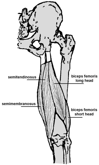
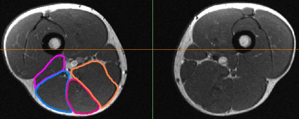
MR images were obtained from 14 athletes who sustained a clinically diagnosed grade I-II hamstring strain injury between 5 and 23 months prior as well as five healthy controls. Qualitative bilateral comparisons were used to assess the presence of fatty infiltration and changes in morphology that may have arisen as a result of the previous injury. Hamstring muscle and tendon-scar volumes were quantified in both limbs for the biceps femoris long head (BFLH), biceps femoris short head (BFSH), the proximal semimembranosus tendon, and the proximal conjoint biceps femoris and semitendinosus tendon. Differences in muscle and tendon volume between limbs were statistically compared between the previously injured and healthy control subjects.
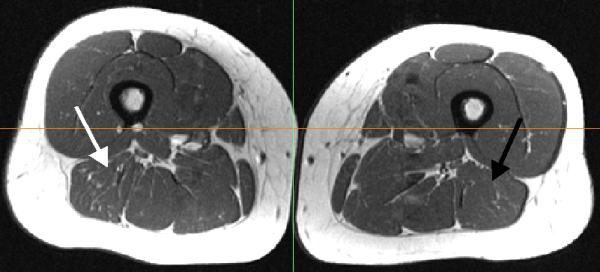
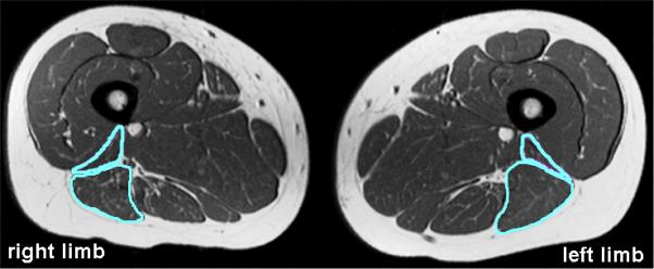
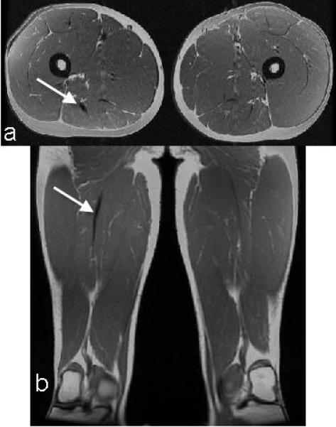
Increased low-intensity signal was present along the musculotendon junction adjacent to the site of presumed prior injury for 11 of the 14 subjects, suggestive of persistent scar tissue. The 13 subjects with biceps femoris injuries displayed a significant decrease in BFLH volume (p < 0.01), often accompanied by an increase in BFSH volume. Two of these subjects also presented with fatty infiltration within the previously injured BFLH.
The results of this study provide evidence of long-term musculotendon remodeling following a hamstring strain injury. Additionally, many athletes are likely returning to sport with residual atrophy of the BFLH and/or hypertrophy of the BFSH. It is possible that long-term changes in musculotendon structure following injury alters contraction mechanics during functional movement, such as running and may contribute to reinjury risk.
本研究的目的是使用磁共振成像来研究腘绳肌拉伤后肌肉和肌腱形态的长期变化。
对14名在5至23个月前临床诊断为I-II级腘绳肌拉伤的运动员以及5名健康对照者进行磁共振成像检查。采用双侧定性比较来评估脂肪浸润的存在以及先前损伤可能导致的形态变化。对双侧的股二头肌长头(BFLH)、股二头肌短头(BFSH)、半膜肌近端肌腱以及股二头肌和半腱肌近端联合肌腱的腘绳肌和肌腱瘢痕体积进行量化。对先前受伤的受试者和健康对照者的肢体间肌肉和肌腱体积差异进行统计学比较。
14名受试者中有11名在假定先前损伤部位附近的肌-腱连接处出现低强度信号增加,提示存在持续的瘢痕组织。13名股二头肌受伤的受试者BFLH体积显著减小(p < 0.01),且常伴有BFSH体积增加。其中两名受试者在先前受伤的BFLH内还出现了脂肪浸润。
本研究结果为腘绳肌拉伤后长期的肌-腱重塑提供了证据。此外,许多运动员可能在BFLH残留萎缩和/或BFSH肥大的情况下重返运动。受伤后肌-腱结构的长期变化可能会改变跑步等功能性运动中的收缩力学,并可能增加再次受伤的风险。