Geft Dael, Schwartzenberg Shmuel, Rogowsky Ori, Finkelstein Ariel, Ablin Jacob, Maysel-Auslender Sofia, Wexler Dov, Keren Gad, George Jacob
Department of Cardiology, Tel Aviv Sourasky Medical Center, Tel Aviv, Israel.
PLoS One. 2008 Sep 18;3(9):e3238. doi: 10.1371/journal.pone.0003238.
Circulating CD34+ endothelial progenitor cells (EPCs) are capable of differentiating into mature endothelial cells to assist in angiogenesis and vasculogenesis. We sought to quantify the numbers of apoptotic progenitors in patients with congestive heart failure.
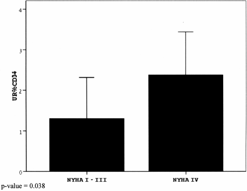
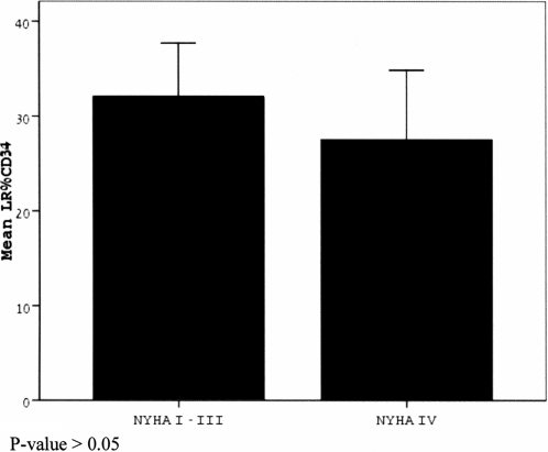
Peripheral blood mononuclear cells were isolated by Ficoll density-gradient from 58 patients with various degrees of heart failure and 23 matched controls. Apoptosis in progenitor CD34+ cells was assessed using the Annexin V-PE/PI detection kit, and FACS analysis was performed with triple staining for CD34, annexin-V and propidium iodide. The percentage of early and late apoptotic progenitor cells was determined in the subject groups and was correlated with clinical characteristics. While there was no significant difference in total CD34 positive cells or early apoptotic progenitors between control subjects and CHF patients (p = 0.42) or between severe and mild/moderate CHF groups (p = 0.544), there was an elevated number of late apoptotic progenitors in the severe CHF group compared with the mild/moderate CHF group (p = 0.03). Late apoptotic progenitors were significantly increased in CHF patients as compared to matched controls. There was also an inverse correlation between late apoptotic progenitors and ejection fraction (r = -0.252, p = 0.028) as well as a positive association with NYHA class (r = 0.223, p = 0.046).
Severe heart failure patients exhibited higher numbers of late apoptotic progenitors, and this was positively associated with NYHA class and negatively correlated with ejection fraction. This finding may shed light on the numerous factors governing the pathophysiology of CHF.
循环中的CD34+内皮祖细胞(EPCs)能够分化为成熟内皮细胞,以协助血管生成和血管发生。我们试图对充血性心力衰竭患者中凋亡祖细胞的数量进行量化。
通过Ficoll密度梯度法从58例不同程度心力衰竭患者和23例匹配对照中分离外周血单个核细胞。使用膜联蛋白V-PE/PI检测试剂盒评估祖细胞CD34+中的凋亡情况,并对CD34、膜联蛋白V和碘化丙啶进行三重染色后进行流式细胞术分析。在各研究组中测定早期和晚期凋亡祖细胞的百分比,并将其与临床特征相关联。对照受试者与CHF患者之间(p = 0.42)或重度与轻度/中度CHF组之间(p = 0.544),总的CD34阳性细胞或早期凋亡祖细胞无显著差异,但重度CHF组与轻度/中度CHF组相比,晚期凋亡祖细胞数量增加(p = 0.03)。与匹配对照相比,CHF患者中晚期凋亡祖细胞显著增加。晚期凋亡祖细胞与射血分数呈负相关(r = -0.252,p = 0.028),与纽约心脏协会(NYHA)心功能分级呈正相关(r = 0.223,p = 0.046)。
重度心力衰竭患者表现出更高数量的晚期凋亡祖细胞,这与NYHA心功能分级呈正相关,与射血分数呈负相关。这一发现可能有助于揭示控制CHF病理生理学的众多因素。