Estler Mary, Boskovic Goran, Denvir James, Miles Sarah, Primerano Donald A, Niles Richard M
Department of Biochemistry and Microbiology, Joan C. Edwards School of Medicine, Marshall University, One John Marshall Drive - BBSC, Huntington, WV 25755, USA.
BMC Genomics. 2008 Oct 11;9:478. doi: 10.1186/1471-2164-9-478.
The incidence of malignant melanoma has significantly increased over the last decade. Some of these malignancies are susceptible to the growth inhibitory and pro-differentiating effects of all-trans-retinoic acid (RA). The molecular changes responsible for the biological activity of RA in melanoma are not well understood.
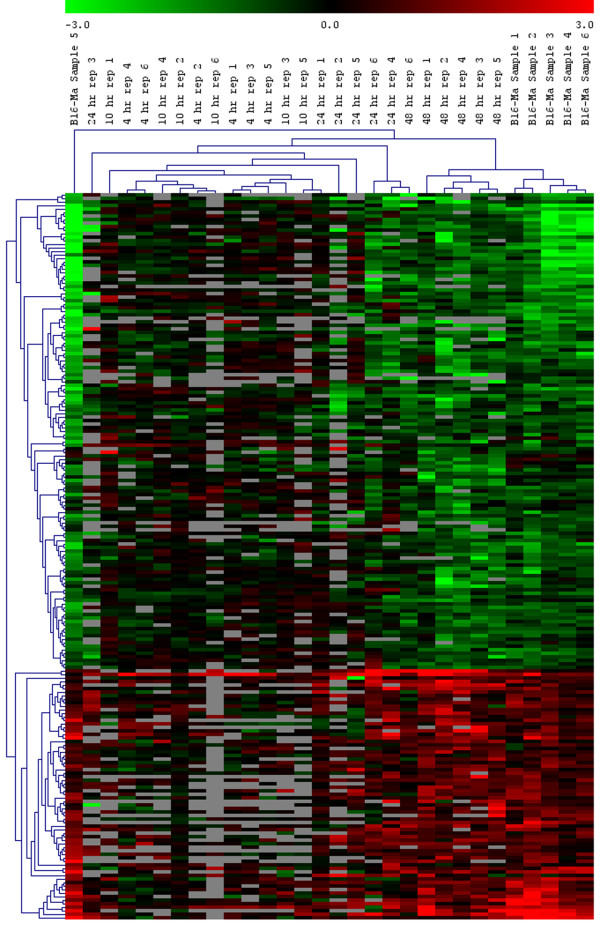
In an analysis of sequential global gene expression changes during a 4-48 h RA treatment of B16 mouse melanoma cells, we found that RA increased the expression of 757 genes and decreased the expression of 737 genes. We also compared the gene expression profile (no RA treatment) between non-malignant melan-a mouse melanocytes and B16 melanoma cells. Using the same statistical test, we found 1495 genes whose expression was significantly higher in melan-a than in B16 cells and 2054 genes whose expression was significantly lower in melan-a than in B16 cells. By intersecting these two gene sets, we discovered a common set of 233 genes whose RNA levels were significantly different between B16 and melan-a cells and whose expression was altered by RA treatment. Within this set, RA treatment altered the expression of 203 (87%) genes toward the melan-a expression level. In addition, hierarchical clustering showed that after 48 h of RA treatment expression of the 203 genes was more closely related to the melan-a gene set than any other RA treatment time point. Functional analysis of the 203 gene set indicated that RA decreased expression of mRNAs that encode proteins involved in cell division/cell cycle, DNA replication, recombination and repair, and transcription regulation. Conversely, it stimulated genes involved in cell-cell signaling, cell adhesion and cell differentiation/embryonic development. Pathway analysis of the 203 gene set revealed four major hubs of connectivity: CDC2, CHEK1, CDC45L and MCM6.
Our analysis of common genes in the 48 h RA-treatment of B16 melanoma cells and untreated B16 vs. melan-a data set show that RA "normalized" the expression of genes involved in energy metabolism, DNA replication, DNA repair and differentiation. These results are compatible with the known growth inhibitory and pro-differentiating effects of RA. Pathway analysis suggests that CDC2, CHEK1, CDC45L and MCM6 are key players in mediating the biological activity of RA in B16 melanoma cells.
在过去十年中,恶性黑色素瘤的发病率显著上升。其中一些恶性肿瘤对全反式维甲酸(RA)的生长抑制和促分化作用敏感。RA在黑色素瘤中发挥生物学活性所涉及的分子变化尚未完全明确。
在对B16小鼠黑色素瘤细胞进行4 - 48小时RA处理过程中的连续全基因组表达变化分析中,我们发现RA使757个基因的表达增加,737个基因的表达减少。我们还比较了非恶性黑色素 - a小鼠黑素细胞和B16黑色素瘤细胞之间的基因表达谱(未进行RA处理)。使用相同的统计检验,我们发现1495个基因在黑色素 - a中的表达显著高于B16细胞,2054个基因在黑色素 - a中的表达显著低于B16细胞。通过交叉这两个基因集,我们发现了一组共233个基因,其RNA水平在B16和黑色素 - a细胞之间存在显著差异,并且其表达受到RA处理的影响。在这个基因集中,RA处理使203个(87%)基因的表达朝着黑色素 - a的表达水平改变。此外,层次聚类显示,在RA处理48小时后,这203个基因的表达与黑色素 - a基因集的相关性比任何其他RA处理时间点都更高。对这203个基因集的功能分析表明,RA降低了编码参与细胞分裂/细胞周期、DNA复制、重组和修复以及转录调控的蛋白质的mRNA的表达。相反,它刺激了参与细胞 - 细胞信号传导、细胞粘附和细胞分化/胚胎发育的基因。对这203个基因集的通路分析揭示了四个主要的连接枢纽:CDC2、CHEK1、CDC45L和MCM6。
我们对B16黑色素瘤细胞48小时RA处理以及未处理的B16与黑色素 - a数据集的共同基因分析表明,RA使参与能量代谢、DNA复制、DNA修复和分化的基因表达“正常化”。这些结果与RA已知的生长抑制和促分化作用相符。通路分析表明,CDC2、CHEK1、CDC45L和MCM6是介导RA在B16黑色素瘤细胞中生物学活性的关键因子。