Xie Weijiao, Matsumoto Misaki, Chun Jerold, Ueda Hiroshi
Division of Molecular Pharmacology and Neuroscience, Nagasaki University Graduate School of Biomedical Sciences, Nagasaki 852-8521, Japan.
Mol Pain. 2008 Oct 15;4:46. doi: 10.1186/1744-8069-4-46.
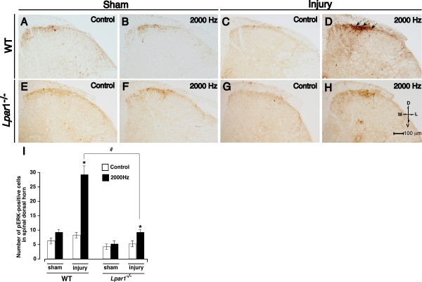
Lysophosphatidic acid receptor subtype LPA(1) is crucial for the initiation of neuropathic pain and underlying changes, such as up-regulation of Ca2+ channel alpha2delta-1 subunit in dorsal root ganglia (DRG), up-regulation of PKCgamma in the spinal dorsal horn, and demyelination of dorsal root fibers. In the present study, we further examined the involvement of LPA(1) signaling in the reorganization of Abeta-fiber-mediated spinal transmission, which is presumed to underlie neuropathic allodynia. Following nerve injury, the phosphorylation of extracellular-signal regulated kinase (pERK) by Abeta-fiber stimulation was observed in the superficial layer of spinal dorsal horn, where nociceptive C- or Adelta-fibers are innervated, but not in sham-operated wild-type mice. However, the pERK signals were largely abolished in LPA(1) receptor knock-out (Lpar1-/-) mice, further supported by quantitative analyses of pERK-positive cells. These results suggest that LPA(1) receptor-mediated signaling mechanisms also participate in functional cross-talk between Abeta- and C- or Adelta-fibers.
溶血磷脂酸受体亚型LPA(1)对于神经性疼痛的起始及潜在变化至关重要,这些变化包括背根神经节(DRG)中Ca2+通道α2δ-1亚基的上调、脊髓背角中PKCγ的上调以及背根纤维的脱髓鞘。在本研究中,我们进一步研究了LPA(1)信号传导在Aβ纤维介导的脊髓传递重组中的作用,这种重组被认为是神经性异常性疼痛的基础。神经损伤后,在脊髓背角浅层观察到Aβ纤维刺激引起的细胞外信号调节激酶(pERK)磷酸化,伤害性C纤维或Aδ纤维在此处支配,但在假手术的野生型小鼠中未观察到。然而,pERK信号在LPA(1)受体敲除(Lpar1-/-)小鼠中基本消失,pERK阳性细胞的定量分析进一步支持了这一结果。这些结果表明,LPA(1)受体介导的信号传导机制也参与了Aβ纤维与C纤维或Aδ纤维之间的功能串扰。