Castro Nadia P, Osório Cynthia A B T, Torres César, Bastos Elen P, Mourão-Neto Mário, Soares Fernando A, Brentani Helena P, Carraro Dirce M
Laboratório de Genômica e Biologia Molecular, Centro de Pesquisa Hospital do Câncer A C Camargo, São Paulo, SP, Brazil.
Breast Cancer Res. 2008;10(5):R87. doi: 10.1186/bcr2157. Epub 2008 Oct 17.
Ductal carcinoma in situ (DCIS) of the breast includes a heterogeneous group of preinvasive tumors with uncertain evolution. Definition of the molecular factors necessary for progression to invasive disease is crucial to determining which lesions are likely to become invasive. To obtain insight into the molecular basis of DCIS, we compared the gene expression pattern of cells from the following samples: non-neoplastic, pure DCIS, in situ component of lesions with co-existing invasive ductal carcinoma, and invasive ductal carcinoma.
Forty-one samples were evaluated: four non-neoplastic, five pure DCIS, 22 in situ component of lesions with co-existing invasive ductal carcinoma, and 10 invasive ductal carcinoma. Pure cell populations were isolated using laser microdissection. Total RNA was purified, DNase treated, and amplified using the T7-based method. Microarray analysis was conducted using a customized cDNA platform. The concept of molecular divergence was applied to classify the sample groups using analysis of variance followed by Tukey's test.
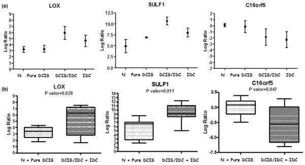
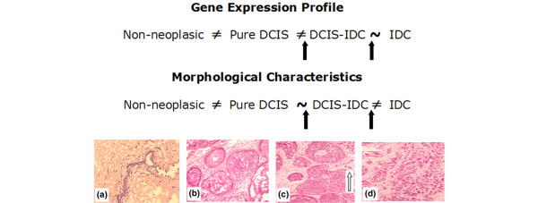
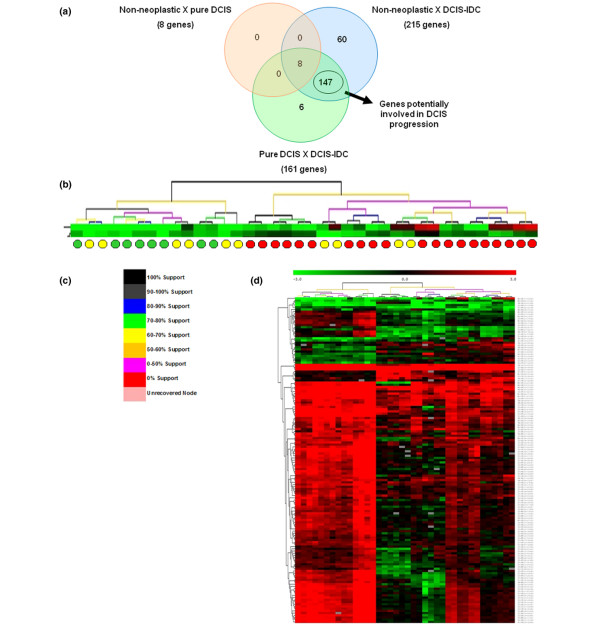
Among the tumor sample groups, cells from pure DCIS exhibited the most divergent molecular profile, consequently identifying cells from in situ component of lesions with co-existing invasive ductal carcinoma as very similar to cells from invasive lesions. Additionally, we identified 147 genes that were differentially expressed between pure DCIS and in situ component of lesions with co-existing invasive ductal carcinoma, which can discriminate samples representative of in situ component of lesions with co-existing invasive ductal carcinoma from 60% of pure DCIS samples. A gene subset was evaluated using quantitative RT-PCR, which confirmed differential expression for 62.5% and 60.0% of them using initial and partial independent sample groups, respectively. Among these genes, LOX and SULF-1 exhibited features that identify them as potential participants in the malignant process of DCIS.
We identified new genes that are potentially involved in the malignant transformation of DCIS, and our findings strongly suggest that cells from the in situ component of lesions with co-existing invasive ductal carcinoma exhibit molecular alterations that enable them to invade the surrounding tissue before morphological changes in the lesion become apparent.
乳腺导管原位癌(DCIS)包括一组异质性的侵袭前肿瘤,其演变情况尚不确定。确定进展为浸润性疾病所需的分子因素对于判断哪些病变可能发展为浸润性病变至关重要。为深入了解DCIS的分子基础,我们比较了以下样本细胞的基因表达模式:非肿瘤性、纯DCIS、伴有浸润性导管癌的病变的原位成分以及浸润性导管癌。
评估了41个样本:4个非肿瘤性样本、5个纯DCIS样本、22个伴有浸润性导管癌的病变的原位成分样本以及10个浸润性导管癌样本。使用激光显微切割分离纯细胞群体。纯化总RNA,进行DNase处理,并使用基于T7的方法进行扩增。使用定制的cDNA平台进行微阵列分析。应用分子差异概念,通过方差分析和Tukey检验对样本组进行分类。
在肿瘤样本组中,纯DCIS的细胞表现出最具差异的分子特征,因此将伴有浸润性导管癌的病变的原位成分的细胞鉴定为与浸润性病变的细胞非常相似。此外,我们鉴定出147个在纯DCIS和伴有浸润性导管癌的病变的原位成分之间差异表达的基因,这些基因能够从60%的纯DCIS样本中区分出代表伴有浸润性导管癌的病变的原位成分的样本。使用定量RT-PCR评估了一个基因子集,分别使用初始和部分独立样本组确认其中62.5%和60.0%的基因存在差异表达。在这些基因中,LOX和SULF-1表现出的特征表明它们可能是DCIS恶性过程的潜在参与者。
我们鉴定出了可能参与DCIS恶性转化的新基因,我们的研究结果强烈表明,伴有浸润性导管癌的病变的原位成分的细胞表现出分子改变,使它们能够在病变形态变化明显之前侵入周围组织。