Wu Jiantao, Gong Jun, Geng Juan, Song Yinxue
Department of Gastroenterology, the Second Affiliated Hospital, Xi'an Jiaotong University School of Medicine, Xi'an, Shaanxi 710004, PR China.
BMC Cancer. 2008 Nov 13;8:333. doi: 10.1186/1471-2407-8-333.
Mucin alterations are a common feature of esophageal neoplasia, and alterations in MUC2 mucin have been associated with tumor progression in the esophagus. Bile acids have been linked to esophageal adenocarcinoma and mucin secretion, but their effects on mucin gene expression in human esophageal adenocarcinoma cells is unknown.
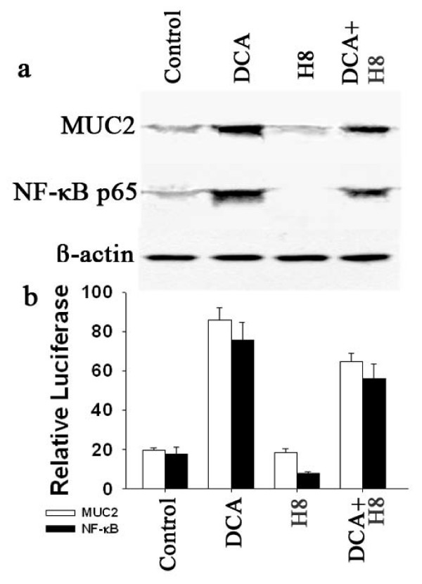
Human esophageal adenocarcinoma cells were treated 18 hours with 50-300 muM deoxycholic acid, chenodeoxycholic acid, or taurocholic acid. MUC2 transcription was assayed using a MUC2 promoter reporter luciferase construct and MUC2 protein was assayed by Western blot analysis. Transcription Nuclear factor-kappaB activity was measured using a Nuclear factor-kappaB reporter construct and confirmed by Western blot analysis for Nuclear factor-kappaB p65.
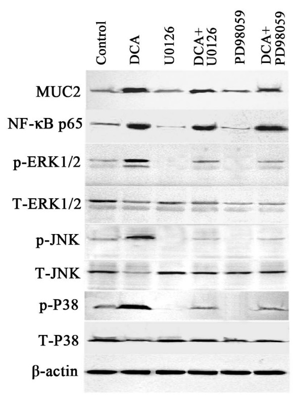
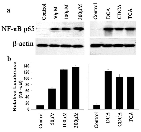
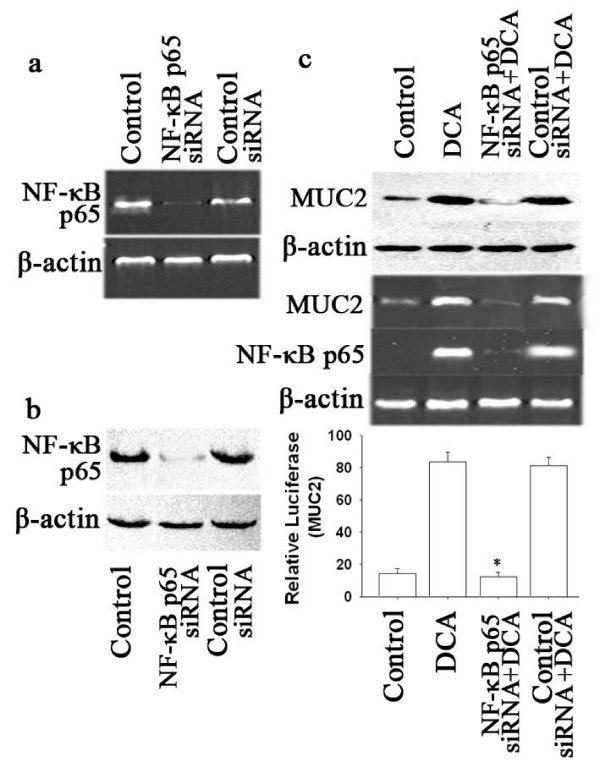
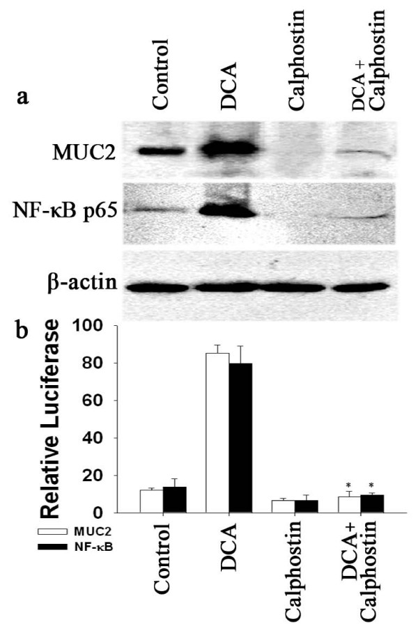
MUC2 transcription and MUC2 protein expression were increased four to five fold by bile acids in a time and dose-dependent manner with no effect on cell viability. Nuclear factor-kappaB activity was also increased. Treatment with the putative chemopreventive agent aspirin, which decreased Nuclear factor-kappaB activity, also decreased MUC2 transcription. Nuclear factor-kappaB p65 siRNA decreased MUC2 transcription, confirming the significance of Nuclear factor-kappaB in MUC2 induction by deoxycholic acid. Calphostin C, a specific inhibitor of protein kinase C (PKC), greatly decreased bile acid induced MUC2 transcription and Nuclear factor-kappaB activity, whereas inhibitors of MAP kinase had no effect.
Deoxycholic acid induced MUC2 overexpression in human esophageal adenocarcinoma cells by activation of Nuclear factor-kappaB transcription through a process involving PKC-dependent but not PKA, independent of activation of MAP kinase.
粘蛋白改变是食管肿瘤形成的常见特征,MUC2粘蛋白的改变与食管肿瘤进展相关。胆汁酸与食管腺癌及粘蛋白分泌有关,但其对人食管腺癌细胞中粘蛋白基因表达的影响尚不清楚。
用50 - 300μM脱氧胆酸、鹅脱氧胆酸或牛磺胆酸处理人食管腺癌细胞18小时。使用MUC2启动子报告荧光素酶构建体检测MUC2转录,通过蛋白质印迹分析检测MUC2蛋白。使用核因子-κB报告构建体测量转录核因子-κB活性,并通过蛋白质印迹分析核因子-κB p65进行确认。
胆汁酸以时间和剂量依赖性方式使MUC2转录和MUC2蛋白表达增加4至5倍,对细胞活力无影响。核因子-κB活性也增加。用假定的化学预防剂阿司匹林处理可降低核因子-κB活性,同时也降低MUC2转录。核因子-κB p65小干扰RNA降低MUC2转录,证实了核因子-κB在脱氧胆酸诱导MUC2表达中的重要性。蛋白激酶C(PKC)的特异性抑制剂钙泊三醇大大降低了胆汁酸诱导的MUC2转录和核因子-κB活性,而丝裂原活化蛋白激酶抑制剂则无作用。
脱氧胆酸通过激活核因子-κB转录诱导人食管腺癌细胞中MUC2过表达,该过程涉及PKC依赖性而非PKA依赖性,且独立于丝裂原活化蛋白激酶的激活。